Copyright
©2013 Baishideng Publishing Group Co.
World J Gastroenterol. Sep 28, 2013; 19(36): 5988-5999
Published online Sep 28, 2013. doi: 10.3748/wjg.v19.i36.5988
Published online Sep 28, 2013. doi: 10.3748/wjg.v19.i36.5988
Figure 1 The scheme for whole experiment.
EA: Electroacupuncture; ST36: Zusanli.
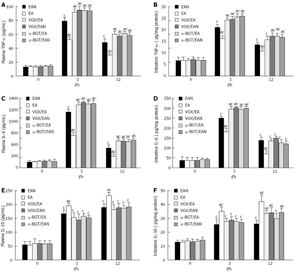
Figure 2 Tumor necrosis factor-α, interleukin-6 and interleukin-10 levels in plasma and intestine at 0, 3 and 12 h after blood loss.
Blood samples and intestine were obtained at 0, 3 and 12 h after blood loss. Data are expressed as mean ± SD (3-5 animals per group at each time point). aP < 0.05 vs EAN group; cP < 0.05 vs 0 h in the same group. EA: Electroacupuncture; VGX: Vagotomy; α-BGT: α-bungarotoxin; TNF: Tumor necrosis factor; IL: Interleukin.
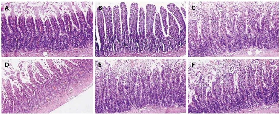
Figure 3 Intestinal histology at 12 h after blood loss.
Electroacupuncture (EA) at ST36 protected against intestinal injury after hemorrhagic shock and delayed fluid replacement, whereas EA at ST36 after vagotomy or injection of α-bungarotoxin eliminated such protection. Sections of distal ileum were harvested at 12 h after blood loss and stained with hematoxylin and eosin. All images are taken at × 200 magnification with black bar = 5 μm (3-5 animals per group at 12 h after blood loss).
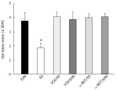
Figure 4 Gut injury scores at 12 h after blood loss.
Gut injury was scored by a pathologist blinded to the experimental groups on a scale of 0-4, (as described in Materials and Methods). aP < 0.05 vs EAN group, (3-5 animals per group at 12 h after blood loss). EA: Electroacupuncture; VGX: Vagotomy; α-BGT: α-bungarotoxin.
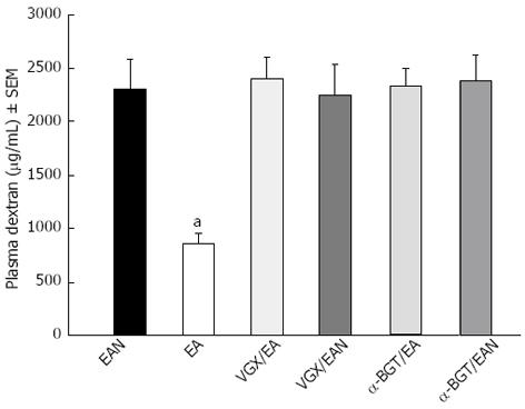
Figure 5 Intestinal permeability to 4-kDa fluorescein isothiocyanate-dextran at 12 h after blood loss.
Electroacupuncture (EA) at ST36 protected the intestine from an increase in permeability after hemorrhagic shock and delayed fluid replacement, whereas EA at ST36 after vagotomy or injection of α-bungarotoxin eliminated such protection. aP < 0.05 vs EAN group, (3-5 animals per group at 12 h after blood loss). EA: Electroacupuncture; VGX: Vagotomy; α-BGT: α-bungarotoxin
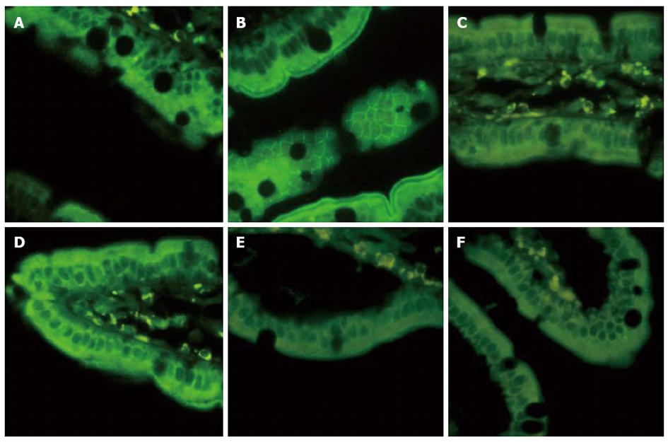
Figure 6 Intestinal ZO-1 immunofluorescent staining at 12 h after blood loss.
Animals in EAN group showed a low fluorescent intensity at the cell periphery after hemorrhagic shock, and electroacupuncture (EA) at ST36 showed preservation of the robust structure of ZO-1 staining, whereas after vagotomy or injection of α-bungarotoxin, it eliminated such protection. All images are taken at × 400 magnification with black bar = 5 μm. (3-5 animals per group at 12 h after blood loss, size bar = 2 μm).
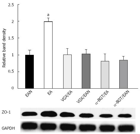
Figure 7 Intestinal ZO-1 protein expression at 12 h after blood loss.
Intestinal extracts were obtained from animals at 12 h after blood loss for measurement of ZO-1 protein expression using Western blotting. Representative Western blotting for the ZO-1 protein is shown with its corresponding glyceraldehyde 3-phosphate dehydrogenase loading control to demonstrate equal protein load in all lanes. Electroacupuncture at ST36 resulted in preservation of protein expression. Significant reduction in ZO-1 expression was seen in all the other groups. aP < 0.05 vs EAN group, (3-5 animals per group at 12h after blood loss). EA: Electroacupuncture; VGX: Vagotomy; α-BGT: α-bungarotoxin; GAPDH: Glyceraldehyde-3-phosphate dehydrogenase.
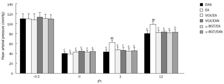
Figure 8 Effect of electroacupuncture ST36 on mean arterial pressure in rats after hemorrhagic shock with delayed fluid replacement.
aP < 0.05 vs EAN group; cP < 0.05 vs 0 h in the same group, (3-5 animals per group at 12 h after blood loss). EA: Electroacupuncture; VGX: Vagotomy; α-BGT: α-bungarotoxin.
- Citation: Du MH, Luo HM, Hu S, Lv Y, Lin ZL, Ma L. Electroacupuncture improves gut barrier dysfunction in prolonged hemorrhagic shock rats through vagus anti-inflammatory mechanism. World J Gastroenterol 2013; 19(36): 5988-5999
- URL: https://www.wjgnet.com/1007-9327/full/v19/i36/5988.htm
- DOI: https://dx.doi.org/10.3748/wjg.v19.i36.5988


