Copyright
©2013 Baishideng Publishing Group Co.
World J Gastroenterol. Sep 28, 2013; 19(36): 5988-5999
Published online Sep 28, 2013. doi: 10.3748/wjg.v19.i36.5988
Published online Sep 28, 2013. doi: 10.3748/wjg.v19.i36.5988
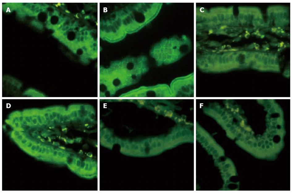
Figure 6 Intestinal ZO-1 immunofluorescent staining at 12 h after blood loss.
Animals in EAN group showed a low fluorescent intensity at the cell periphery after hemorrhagic shock, and electroacupuncture (EA) at ST36 showed preservation of the robust structure of ZO-1 staining, whereas after vagotomy or injection of α-bungarotoxin, it eliminated such protection. All images are taken at × 400 magnification with black bar = 5 μm. (3-5 animals per group at 12 h after blood loss, size bar = 2 μm).
- Citation: Du MH, Luo HM, Hu S, Lv Y, Lin ZL, Ma L. Electroacupuncture improves gut barrier dysfunction in prolonged hemorrhagic shock rats through vagus anti-inflammatory mechanism. World J Gastroenterol 2013; 19(36): 5988-5999
- URL: https://www.wjgnet.com/1007-9327/full/v19/i36/5988.htm
- DOI: https://dx.doi.org/10.3748/wjg.v19.i36.5988


