Copyright
©2013 Baishideng Publishing Group Co.
World J Gastroenterol. Jul 14, 2013; 19(26): 4262-4266
Published online Jul 14, 2013. doi: 10.3748/wjg.v19.i26.4262
Published online Jul 14, 2013. doi: 10.3748/wjg.v19.i26.4262
Figure 1 A white opaque substance-positive gastric hyperplastic polyp is shown on upper endoscopy.
A: An endoscopic examination with a white light image revealed a 25-mm polypoid lesion on the greater curvature in the lower third of the stomach; B: A white opaque substance (WOS) was visualized on the surface of this lesion using conventional endoscopy and magnifying endoscopy (ME) with narrow band imaging (NBI); C: The ME with NBI findings. A regular dotted pattern of the WOS was distributed symmetrically (arrow); D: The ME with NBI findings. An irregular speckled and linear pattern was distributed asymmetrically (arrow).
Figure 2 The resected specimen shows a gastric hyperplastic polyp with dysplasia.
A-D: The histological examination of the resected specimens (hematoxylin and eosin stain). A: In the low power view, the findings for the entire lesion were typical of a gastric hyperplastic polyp; B, C: High-grade dysplasia was observed on the surface of the lesion; D: Low-grade dysplasia was observed on the surface of the lesion; E-I: The immunohistochemical examination. E: Mucin 2 (MUC2); F: MUC5AC; G: MUC6; H: CD10; I: Villin. The lesion had diffuse positivity for MUC5AC, focal positivity for MUC2 and villin, and negative staining for MUC6 and CD10.
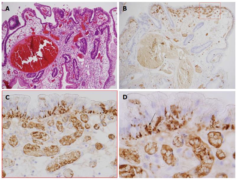
Figure 3 The immunohistochemical analysis indicates that dysplasia is positive for adipophilin.
A: Low-grade dysplasia was observed on the surface of the lesion; B-D: The immunohistochemical examination of adipophilin. Adipophilin was detected in approximately all of the neoplastic cells, especially in the surface epithelium of the intervening apical parts and was located in the subnuclear cytoplasm of the neoplastic cells (arrow).
- Citation: Ueyama H, Matsumoto K, Nagahara A, Gushima R, Hayashi T, Yao T, Watanabe S. A white opaque substance-positive gastric hyperplastic polyp with dysplasia. World J Gastroenterol 2013; 19(26): 4262-4266
- URL: https://www.wjgnet.com/1007-9327/full/v19/i26/4262.htm
- DOI: https://dx.doi.org/10.3748/wjg.v19.i26.4262


