Copyright
©2013 Baishideng Publishing Group Co.
World J Gastroenterol. Jul 14, 2013; 19(26): 4262-4266
Published online Jul 14, 2013. doi: 10.3748/wjg.v19.i26.4262
Published online Jul 14, 2013. doi: 10.3748/wjg.v19.i26.4262
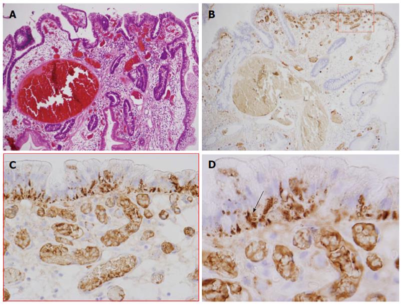
Figure 3 The immunohistochemical analysis indicates that dysplasia is positive for adipophilin.
A: Low-grade dysplasia was observed on the surface of the lesion; B-D: The immunohistochemical examination of adipophilin. Adipophilin was detected in approximately all of the neoplastic cells, especially in the surface epithelium of the intervening apical parts and was located in the subnuclear cytoplasm of the neoplastic cells (arrow).
- Citation: Ueyama H, Matsumoto K, Nagahara A, Gushima R, Hayashi T, Yao T, Watanabe S. A white opaque substance-positive gastric hyperplastic polyp with dysplasia. World J Gastroenterol 2013; 19(26): 4262-4266
- URL: https://www.wjgnet.com/1007-9327/full/v19/i26/4262.htm
- DOI: https://dx.doi.org/10.3748/wjg.v19.i26.4262


