Copyright
©2012 Baishideng Publishing Group Co.
World J Gastroenterol. Feb 28, 2012; 18(8): 778-784
Published online Feb 28, 2012. doi: 10.3748/wjg.v18.i8.778
Published online Feb 28, 2012. doi: 10.3748/wjg.v18.i8.778
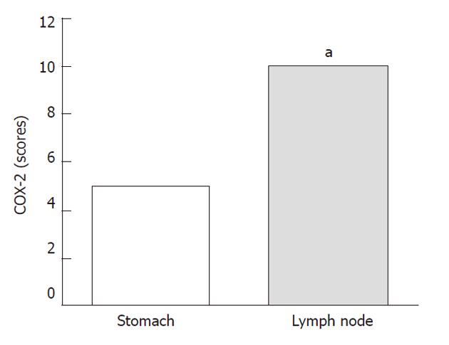
Figure 1 Cyclooxygenase-2 expression in gastric cancer, diffuse histotype.
Lymph node expression is significantly higher than in the stomach (Median scores). aP = 0.0108, Mann-Whitney test. COX-2: Cyclooxygenase-2.
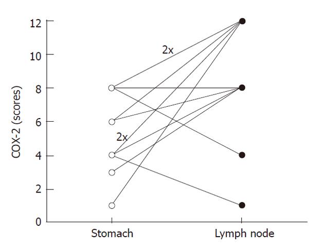
Figure 2 Cyclooxygenase-2 expression in primary diffuse gastric cancer and respective metastasis.
Lymph node expression is higher than stomach expression levels in most cases (Individual case scores). P = 0.0137, Wilcoxon paired test. 2x means two overlaid cases. COX-2: Cyclooxygenase-2.
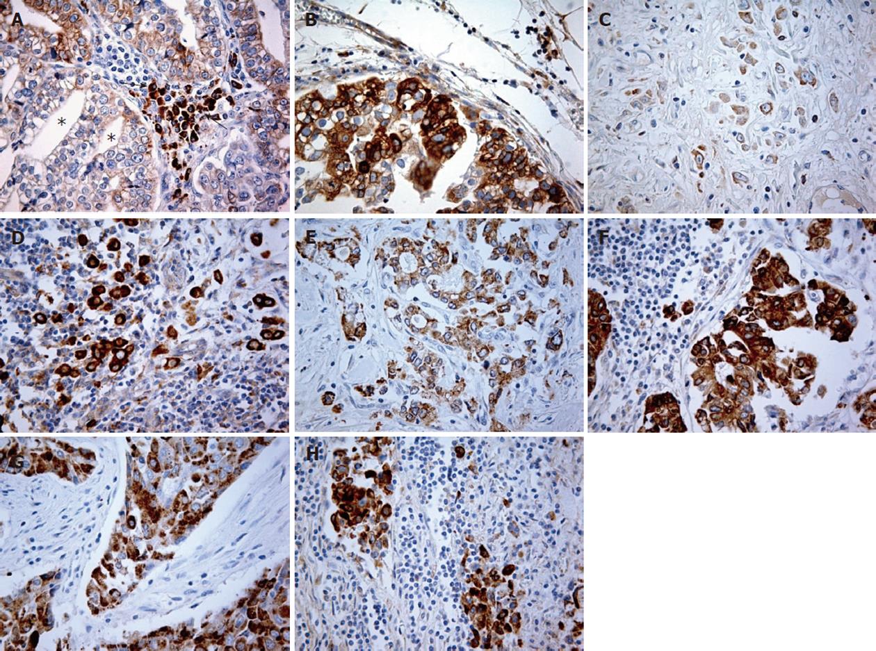
Figure 3 Cyclooxygenase-2 expression in gastric cancer.
A, B: Intestinal type, case 12; A: Stomach = score 1, cancer cells were negative or scarcely stained (*). Strong cyclooxygenase-2 staining of inflammatory cells (mononuclear) into the stroma (arrows); B: Lymph node = score 8; C, D: Diffuse type, case 3; C: Stomach = score 4; D: Lymph node = score 12; E, F: Mixed type, case 7; E: Stomach = score 8 (both components); F: Lymph node = score 12; G, H: Unclassified, case 5; G: Stomach = score 6; H: Lymph node = score 8. All magnifications 400 ×.
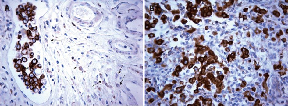
Figure 4 Cyclooxygenase-2 expression in gastric cancer and diffuse type: primary tumor, emboli and metastasis.
A: Stomach: Low cyclooxygenase-2 (COX-2) expression in perivascular malignant cells (arrows). Tumor embolus with strongly stained neoplastic cells (*); B: Lymph node: High COX-2 expression at the same intensity seen in the embolus. Case 3, diffuse histotype, 400 ×.
- Citation: Almeida PR, Ferreira FV, Santos CC, Rocha-Filho FD, Feitosa RR, Falcão EA, Cavada BK, Lima-Júnior RC, Ribeiro RA. Immunoexpression of cyclooxygenase-2 in primary gastric carcinomas and lymph node metastases. World J Gastroenterol 2012; 18(8): 778-784
- URL: https://www.wjgnet.com/1007-9327/full/v18/i8/778.htm
- DOI: https://dx.doi.org/10.3748/wjg.v18.i8.778


