Copyright
©2012 Baishideng Publishing Group Co.
World J Gastroenterol. Apr 14, 2012; 18(14): 1590-1601
Published online Apr 14, 2012. doi: 10.3748/wjg.v18.i14.1590
Published online Apr 14, 2012. doi: 10.3748/wjg.v18.i14.1590
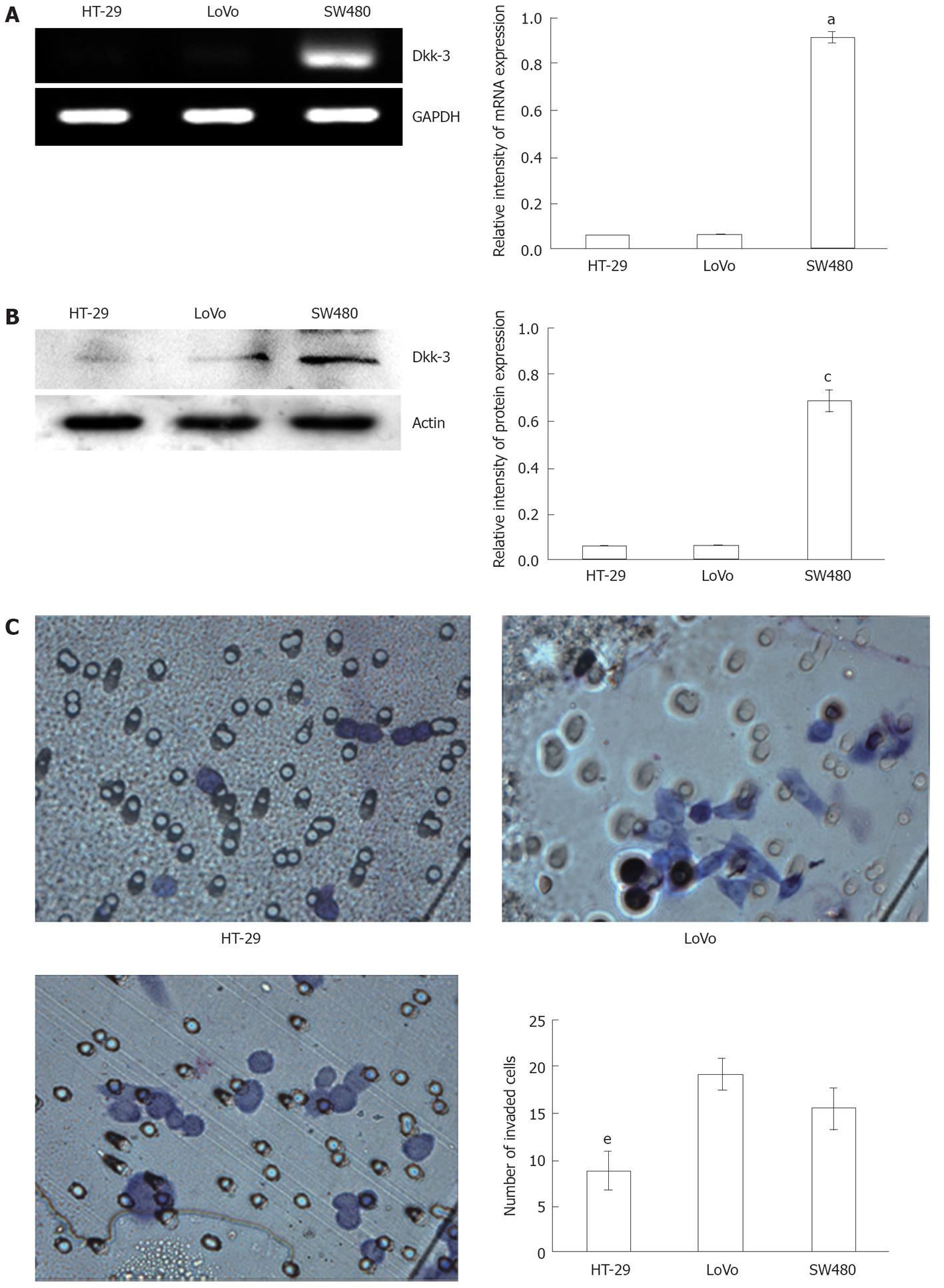
Figure 1 Levels of Dickkopf-3 mRNA and protein expression correlate with invasive potential of human colon cancer cell lines.
A: Semi-quantitative reverse transcription polymerase chain reaction of RNA extracted from colon cancer cell lines, HT-29, LoVo and SW480, respectively, and the glyceraldehyde-3-phosphate dehydrogenase (GAPDH) mRNA was amplified as a control. aP < 0.05 vs HT-29 or LoVo using Student’s t test; B: Endogenous Dickkopf-3 (Dkk-3) protein expression was examined by immunoblot analysis of total cellular protein isolated from three colon cancer cell lines: HT-29, LoVo and SW480, and actin was utilized as a loading control. cP < 0.05 vs HT-29 or LoVo using Student’s t test; C: Human colon cancer cells, HT-29, LoVo and SW480 invading through the Matrigel were counted under a microscope in ten random fields at × 400 magnification. eP < 0.05 vs LoVo or SW480 using Student’s t test.
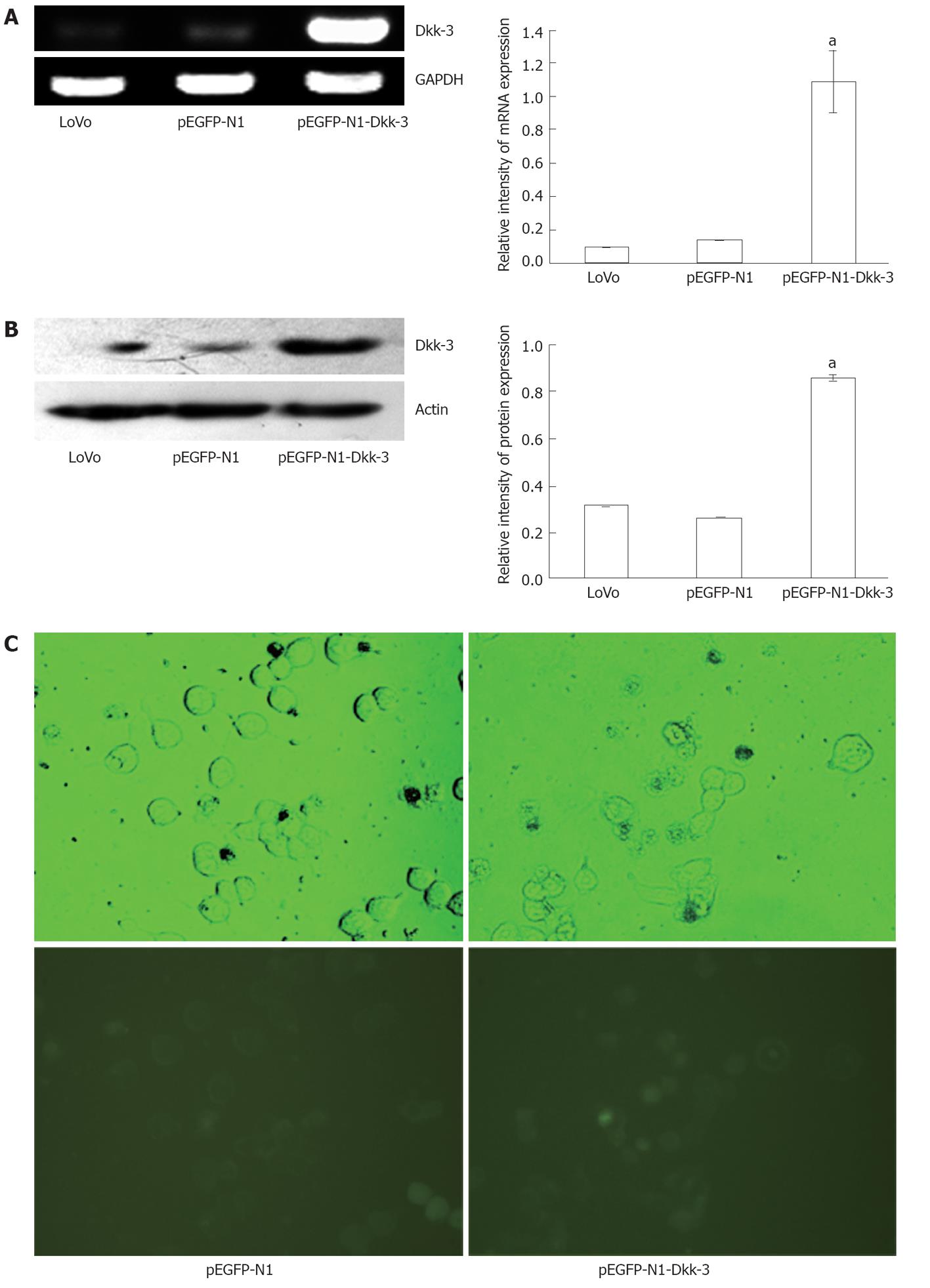
Figure 2 pEGFP-N1-Dkk-3-GFP plasmid induces overexpression of Dickkopf-3 in human colon cancer LoVo cells.
A: Semi-quantitative reverse transcription polymerase chain reaction of RNA extracted from pEGFP-N1-Dkk-3-GFP plasmid transfected LoVo cells and the glyceraldehyde-3-phosphate dehydrogenase (GAPDH) mRNA was amplified as a control; B: Immunoblotting of total protein lysates extracted from pEGFP-N1-Dkk-3-GFP plasmid transfected LoVo cells, and actin was included as a loading control; C: Green fluorescent protein (GFP) was also detected under a fluorescence microscope in pEGFP-N1-Dkk-3-GFP plasmid transfected LoVo cells (× 400). aP < 0.05 vs LoVo or pEGFP-N1 using Student’s t test.
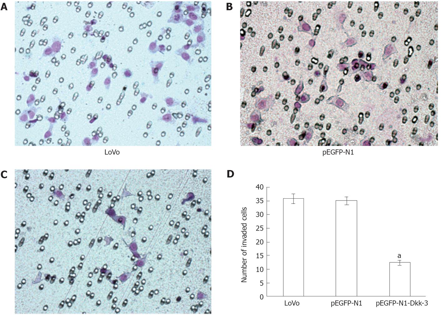
Figure 3 Overexpression of Dickkopf-3 inhibits invasion in human colon cancer LoVo cells.
Representative number of invading cells through the Matrigel was counted under microscope in ten random fields at × 400 magnification. Each bar represented the mean ± SD. aP < 0.05 vs LoVo or pEGFP-N1 using Student’s t test. The results are representative of three separate experiments.
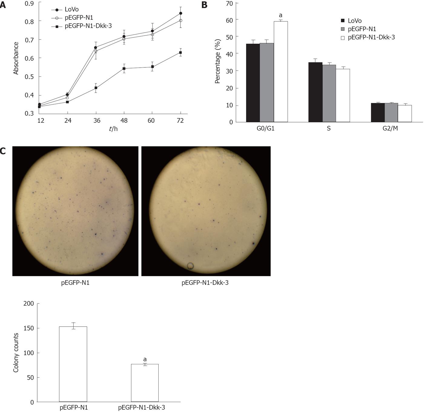
Figure 4 Overexpression of Dickkopf-3 inhibits proliferation and induces G0/G1 arrest in human colon cancer LoVo cells.
A: Dickkopf-3 (Dkk-3) inhibits proliferation in human colon cancer LoVo cells; B: Forty-eight hours after transfection, LoVo was used for cell cycle analysis using a FACSCalibur flow cytometer; C: The dickkopf homolog 3 gene Dkk-3 inhibited tumor cell colony formation. LoVo cells were transfected with pEGFP-N1-Dkk-3-GFP plasmid or with pEGFP-N1 and were maintained in the presence of G418 sulfate for 2 wk. Quantitative analysis of colony numbers are shown as the mean ± SD. aP < 0.05 vs pEGFP-N1 using Student’s t test.
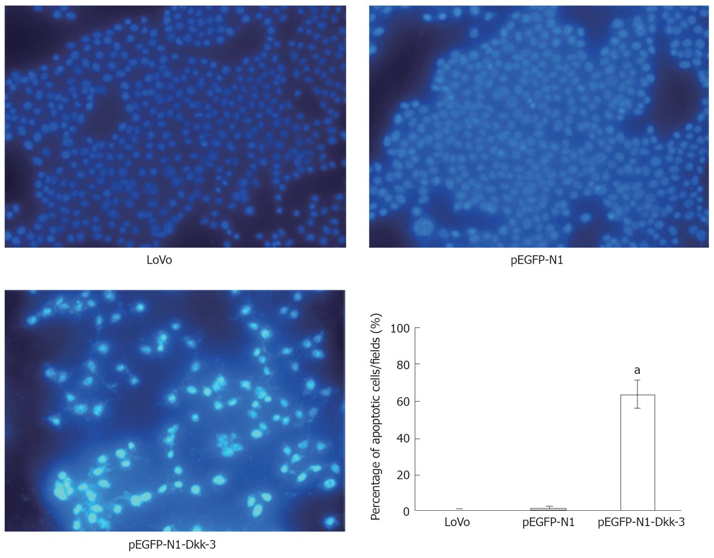
Figure 5 Detection of apoptosis by Hoechst 33285.
The apoptotic feature was assessed by observing chromatin condensation and fragment staining. aP < 0.05 vs control cells (untreated LoVo cells or pEGFP-N1-transfected LoVo cells).
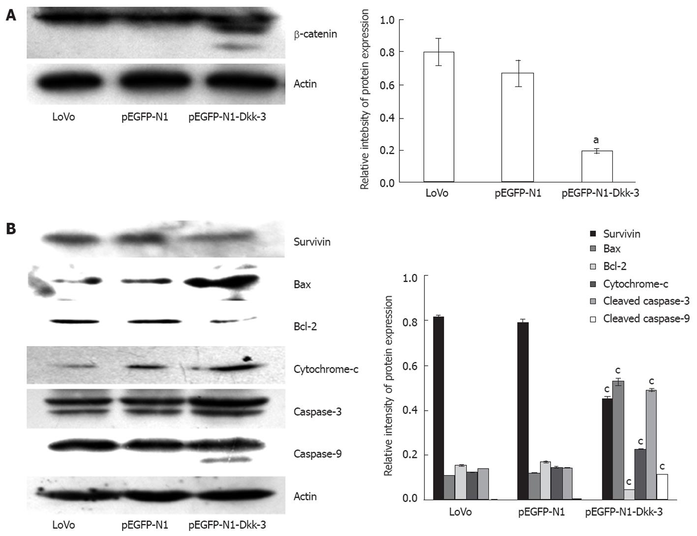
Figure 6 Overexpression of Dickkopf-3 inhibits downstream signaling and induces apoptosis in LoVo cells through mitochondrial pathway.
A: Dickkopf-3 (Dkk-3) reduces cytoplasmic accumulation of β-catenin; B: Western blotting analysis of survivin, Bcl-2, Bax, cytochrome c, caspase-3 and caspase-9 protein after 48 h transfection with pEGFP-N1-Dkk-3-GFP plasmid in LoVo cells. Actin was used as a loading control. aP < 0.05 vs LoVo or pEGFP-N1 using Student’s t test.
- Citation: Yang ZR, Dong WG, Lei XF, Liu M, Liu QS. Overexpression of Dickkopf-3 induces apoptosis through mitochondrial pathway in human colon cancer. World J Gastroenterol 2012; 18(14): 1590-1601
- URL: https://www.wjgnet.com/1007-9327/full/v18/i14/1590.htm
- DOI: https://dx.doi.org/10.3748/wjg.v18.i14.1590


