Copyright
©2012 Baishideng Publishing Group Co.
World J Gastroenterol. Apr 14, 2012; 18(14): 1590-1601
Published online Apr 14, 2012. doi: 10.3748/wjg.v18.i14.1590
Published online Apr 14, 2012. doi: 10.3748/wjg.v18.i14.1590
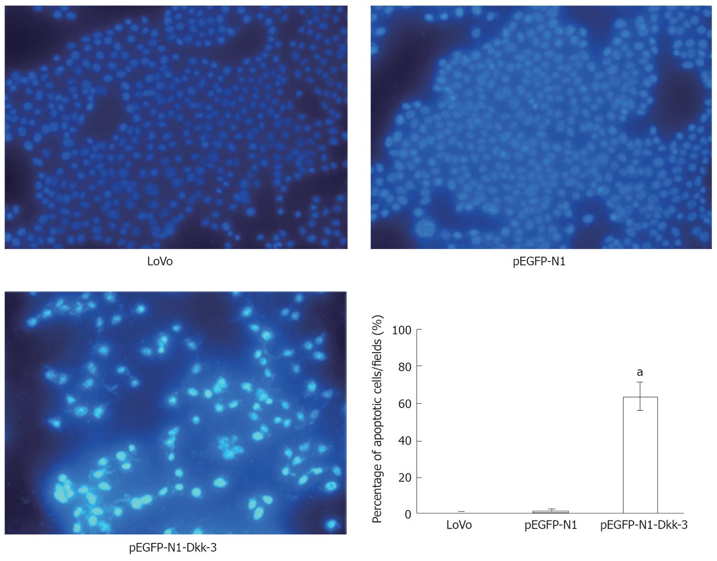
Figure 5 Detection of apoptosis by Hoechst 33285.
The apoptotic feature was assessed by observing chromatin condensation and fragment staining. aP < 0.05 vs control cells (untreated LoVo cells or pEGFP-N1-transfected LoVo cells).
- Citation: Yang ZR, Dong WG, Lei XF, Liu M, Liu QS. Overexpression of Dickkopf-3 induces apoptosis through mitochondrial pathway in human colon cancer. World J Gastroenterol 2012; 18(14): 1590-1601
- URL: https://www.wjgnet.com/1007-9327/full/v18/i14/1590.htm
- DOI: https://dx.doi.org/10.3748/wjg.v18.i14.1590


