Copyright
©The Author(s) 2005.
World J Gastroenterol. Sep 7, 2005; 11(33): 5129-5135
Published online Sep 7, 2005. doi: 10.3748/wjg.v11.i33.5129
Published online Sep 7, 2005. doi: 10.3748/wjg.v11.i33.5129
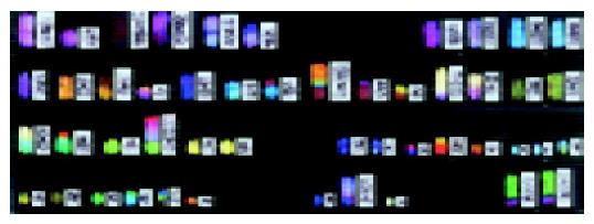
Figure 1 SKY analysis in GT3TKB.
Color images are shown alongside inverted DAPI banding images.
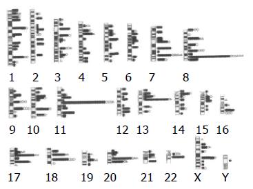
Figure 2 Assignment of breaks in 7 patients with GC and 10 established cell lines.
A total of 630 breaks were identified by SKY. Closed and open circles represent diffuse type and intestinal type, respectively. Open triangle represents the breakpoints obtained from one cell line MKN1 established from a GC patient with adenosquamous cell carcinoma. Mapping analysis of breaks to the ideograms delineated two outstanding loci, 8q24.1 and 11q13. The other breakpoints highlighted are 13q14, 17q11.2, 18q21, and 20q11.2.
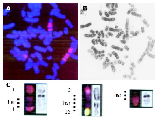
Figure 3 FISH and SKY analysis on SNU16.
A: DC-FISH using YAC probe I2 (8q24.1, red) and cosmid probe CPP29 (11q13.3, green). I2 probe shows periodic staining pattern on HSRs, indicating amplification of MYC gene on HSRs but not double minute chromosomes (DMs). Three copies of CCDN1 gene are detected; B: Inverted DAPI band image of the same metaphase plate shown in A; C: SKY findings of HSRs found in SNU16. Three HSRs were initially identified as originating from chromosome no. 2 based on their fluorescence color.
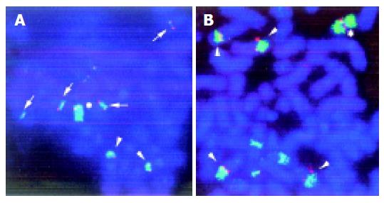
Figure 4 FISH analysis on KMK2.
A: DC-FISH using YAC probe I2 (8q24.1, red) and WCP probe for chromosome 8 (green) demonstrates multiple insertions (arrows) and translocations (arrowheads) of MYC locus. Asterisk indicates a normal chromosome 8. B: DC-FISH using cosmid probe CPP29 (red) and WCP probe for chromosome 11 (green) demonstrates multiple translocations (arrowheads) at CCND1 locus. Asterisk indicates a normal chromosome 11.
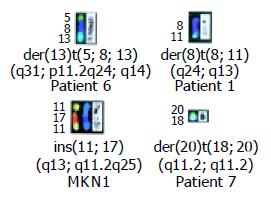
Figure 5 Partial karyotypes of recurrent translocations.
- Citation: Yamashita Y, Nishida K, Okuda T, Nomura K, Matsumoto Y, Mitsufuji S, Horiike S, Hata H, Sakakura C, Hagiwara A, Yamagishi H, Taniwaki M. Recurrent chromosomal rearrangements at bands 8q24 and 11q13 in gastric cancer as detected by multicolor spectral karyotyping. World J Gastroenterol 2005; 11(33): 5129-5135
- URL: https://www.wjgnet.com/1007-9327/full/v11/i33/5129.htm
- DOI: https://dx.doi.org/10.3748/wjg.v11.i33.5129


