Copyright
©The Author(s) 2022.
World J Clin Cases. Oct 26, 2022; 10(30): 11190-11197
Published online Oct 26, 2022. doi: 10.12998/wjcc.v10.i30.11190
Published online Oct 26, 2022. doi: 10.12998/wjcc.v10.i30.11190
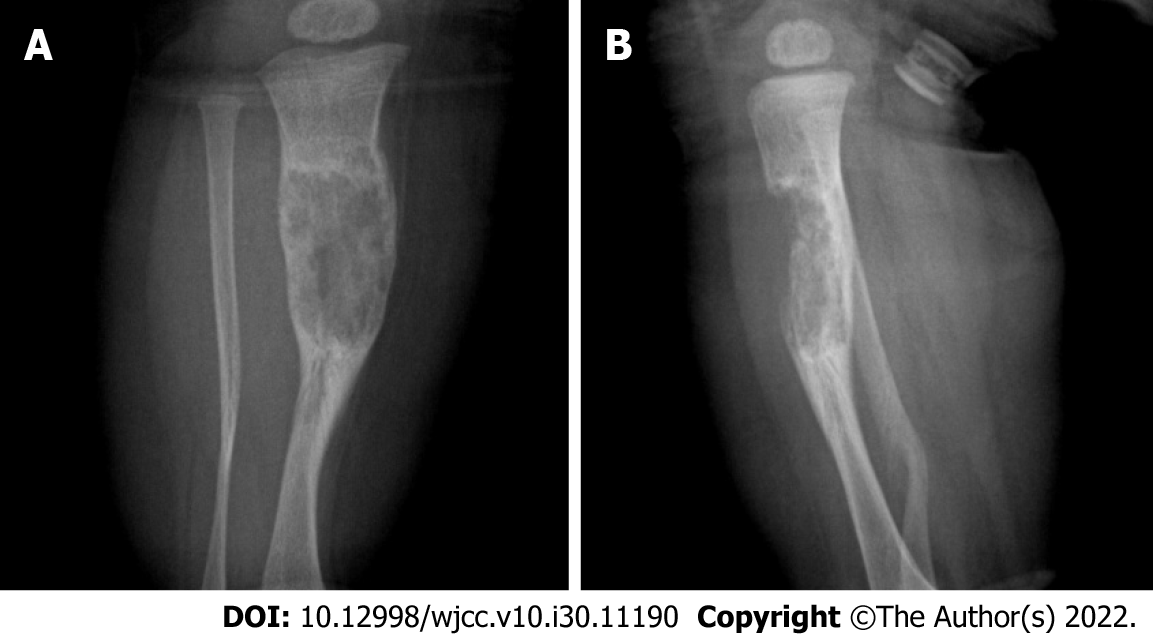
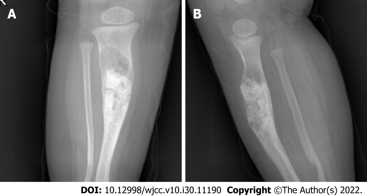
Figure 1 Radiographic examination.
A: Anteroposterior radiographs; B: Lateral radiographs. An area of bone destruction can be seen in the middle part of the right tibia, with uneven internal density, unclear boundaries, slightly expansive, and an anterior cortical bone defect.
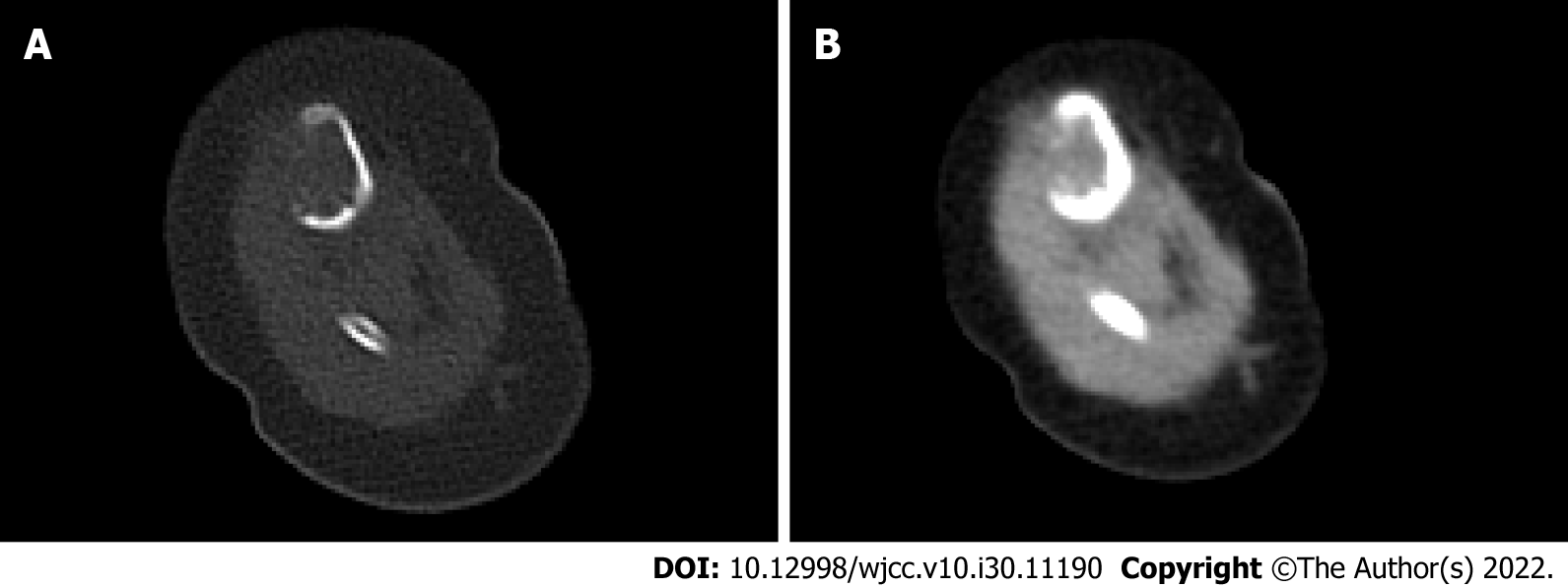
Figure 2 Computed tomography scan.
A: Window of bone tissue; B: Window of soft tissue. Eccentric, expansive soft tissue density shadow in the middle and upper segment of the right tibia, corresponding bone resorption and destruction, and local bone cortical defect.
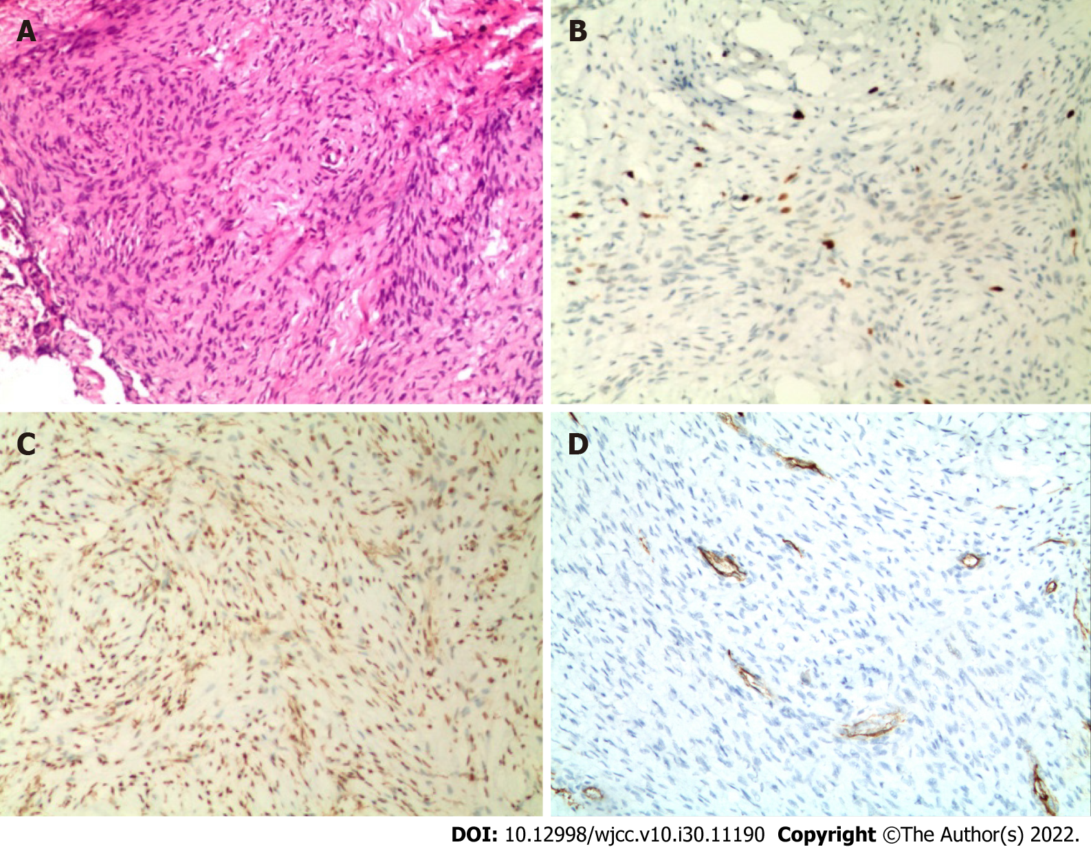
Figure 3 Histopathological examination.
A: Spindle fibroblasts and myofibroblasts arranged in bundles between collagen fibers, spindle or wave nuclei (hematoxylin and eosin stain ×100); B: Immunohistochemical Ki67 (index approximately 5%) (×100); C: Immunochemical smooth muscle actin positive (×100); D: Immunohistochemical CD34 positive (×100).
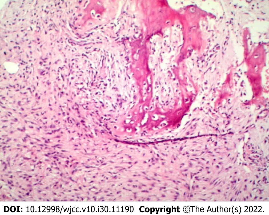
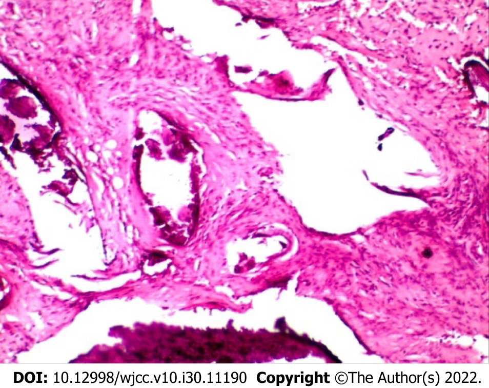
Figure 4 Histopathological examination.
The nuclei are hyperchromatic, star-shaped, ovoid, and wavy, and dense bundles of staggered fibroblasts, among immature bone trabeculae, are seen (×100).
Figure 5 Radiograph.
A: Anteroposterior radiographs; B: Lateral radiographs. Irregular localized bone shape of the middle and upper segment of the right tibia, with uneven internal density and bone graft shadow, with an increase in the size of the original lesion. Irregular and curved bone of the distal segment of the right fibula.
Figure 6 Histopathological examination.
Collagen fibers, adipose tissue, bone tissue, fibroblasts, and myofibroblasts (×100).
- Citation: Qiao YJ, Yang WB, Chang YF, Zhang HQ, Yu XY, Zhou SH, Yang YY, Zhang LD. Fibrous hamartoma of infancy with bone destruction of the tibia: A case report. World J Clin Cases 2022; 10(30): 11190-11197
- URL: https://www.wjgnet.com/2307-8960/full/v10/i30/11190.htm
- DOI: https://dx.doi.org/10.12998/wjcc.v10.i30.11190


