Copyright
©The Author(s) 2022.
World J Clin Cases. Oct 26, 2022; 10(30): 11190-11197
Published online Oct 26, 2022. doi: 10.12998/wjcc.v10.i30.11190
Published online Oct 26, 2022. doi: 10.12998/wjcc.v10.i30.11190
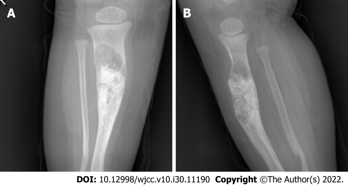
Figure 5 Radiograph.
A: Anteroposterior radiographs; B: Lateral radiographs. Irregular localized bone shape of the middle and upper segment of the right tibia, with uneven internal density and bone graft shadow, with an increase in the size of the original lesion. Irregular and curved bone of the distal segment of the right fibula.
- Citation: Qiao YJ, Yang WB, Chang YF, Zhang HQ, Yu XY, Zhou SH, Yang YY, Zhang LD. Fibrous hamartoma of infancy with bone destruction of the tibia: A case report. World J Clin Cases 2022; 10(30): 11190-11197
- URL: https://www.wjgnet.com/2307-8960/full/v10/i30/11190.htm
- DOI: https://dx.doi.org/10.12998/wjcc.v10.i30.11190


