Copyright
©The Author(s) 2022.
World J Clin Cases. Oct 26, 2022; 10(30): 11190-11197
Published online Oct 26, 2022. doi: 10.12998/wjcc.v10.i30.11190
Published online Oct 26, 2022. doi: 10.12998/wjcc.v10.i30.11190
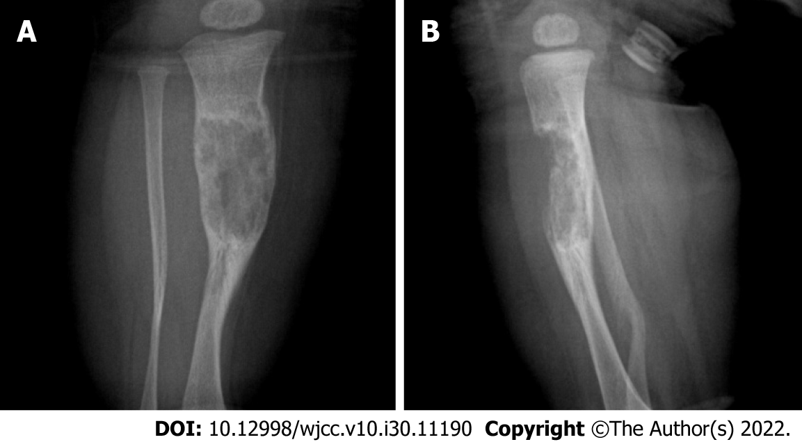
Figure 1 Radiographic examination.
A: Anteroposterior radiographs; B: Lateral radiographs. An area of bone destruction can be seen in the middle part of the right tibia, with uneven internal density, unclear boundaries, slightly expansive, and an anterior cortical bone defect.
- Citation: Qiao YJ, Yang WB, Chang YF, Zhang HQ, Yu XY, Zhou SH, Yang YY, Zhang LD. Fibrous hamartoma of infancy with bone destruction of the tibia: A case report. World J Clin Cases 2022; 10(30): 11190-11197
- URL: https://www.wjgnet.com/2307-8960/full/v10/i30/11190.htm
- DOI: https://dx.doi.org/10.12998/wjcc.v10.i30.11190


