Copyright
©The Author(s) 2022.
World J Clin Cases. Oct 26, 2022; 10(30): 11190-11197
Published online Oct 26, 2022. doi: 10.12998/wjcc.v10.i30.11190
Published online Oct 26, 2022. doi: 10.12998/wjcc.v10.i30.11190
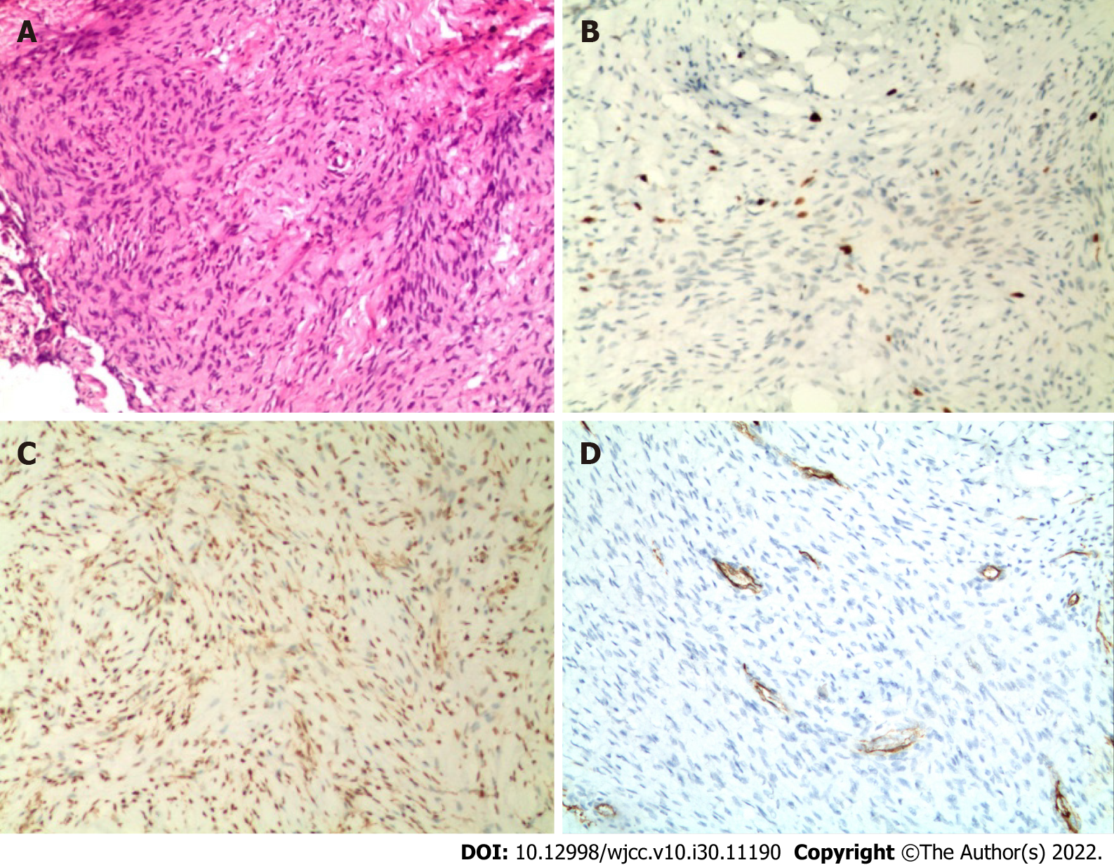
Figure 3 Histopathological examination.
A: Spindle fibroblasts and myofibroblasts arranged in bundles between collagen fibers, spindle or wave nuclei (hematoxylin and eosin stain ×100); B: Immunohistochemical Ki67 (index approximately 5%) (×100); C: Immunochemical smooth muscle actin positive (×100); D: Immunohistochemical CD34 positive (×100).
- Citation: Qiao YJ, Yang WB, Chang YF, Zhang HQ, Yu XY, Zhou SH, Yang YY, Zhang LD. Fibrous hamartoma of infancy with bone destruction of the tibia: A case report. World J Clin Cases 2022; 10(30): 11190-11197
- URL: https://www.wjgnet.com/2307-8960/full/v10/i30/11190.htm
- DOI: https://dx.doi.org/10.12998/wjcc.v10.i30.11190


