Copyright
©2014 Baishideng Publishing Group Inc.
World J Gastroenterol. Jul 14, 2014; 20(26): 8471-8481
Published online Jul 14, 2014. doi: 10.3748/wjg.v20.i26.8471
Published online Jul 14, 2014. doi: 10.3748/wjg.v20.i26.8471
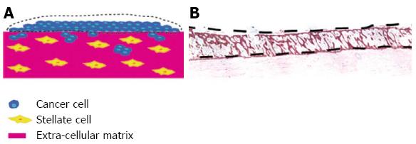
Figure 5 Use of organotypic model to isolate cell types grown together by laser microdisection.
By using the raised organotypic model with pancreatic stellate cells embedded within the gel (Figure 4B) the sellate and cancer cells are kept separate. A, B: Laser microdissection of the cancer cells can then be performed to allow analysis of cancer cells grown in the presence of pancreatic stellate cells. Scale bar 100 μm.
- Citation: Coleman SJ, Watt J, Arumugam P, Solaini L, Carapuca E, Ghallab M, Grose RP, Kocher HM. Pancreatic cancer organotypics: High throughput, preclinical models for pharmacological agent evaluation. World J Gastroenterol 2014; 20(26): 8471-8481
- URL: https://www.wjgnet.com/1007-9327/full/v20/i26/8471.htm
- DOI: https://dx.doi.org/10.3748/wjg.v20.i26.8471


