Copyright
©2013 Baishideng Publishing Group Co.
World J Gastroenterol. Nov 28, 2013; 19(44): 8141-8145
Published online Nov 28, 2013. doi: 10.3748/wjg.v19.i44.8141
Published online Nov 28, 2013. doi: 10.3748/wjg.v19.i44.8141
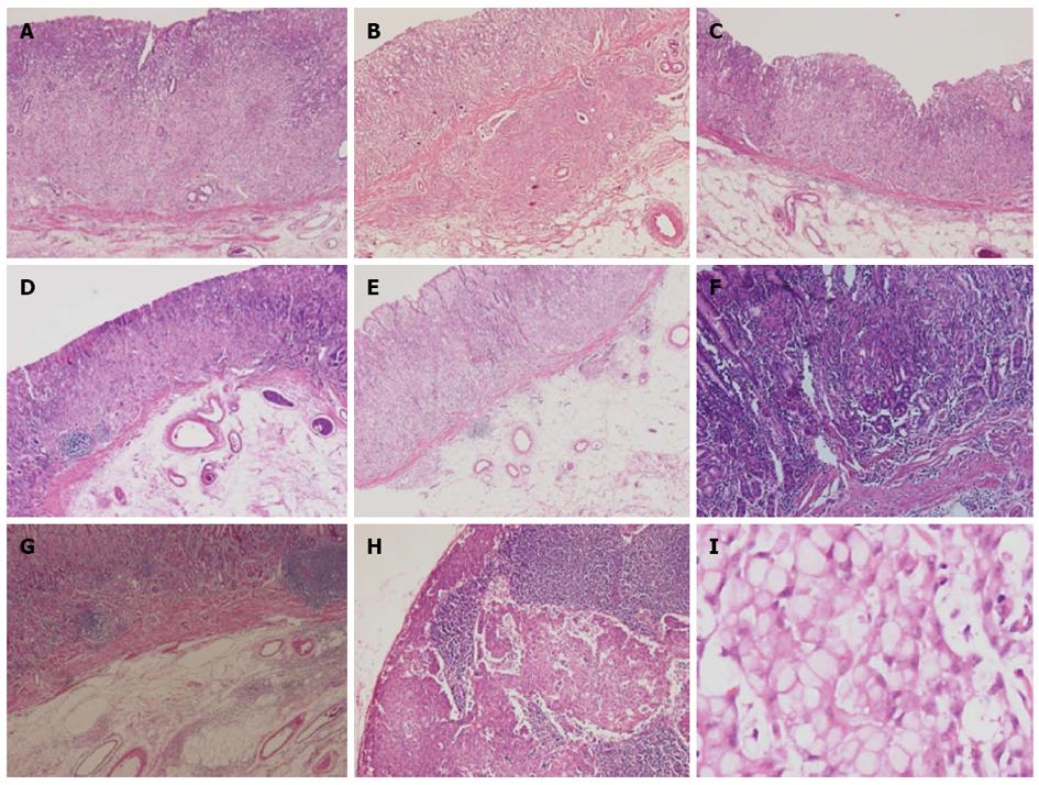
Figure 3 Histopathlogical findings.
A-G: Adenocarcinoma, poorly differentiated in each early gastric cancer lesions in Figure 1 (× 40); H: Lymph node metastasis after gastrectomy (× 40); I: Signet ring cell type of biopsy specimen in esophagogastroduodenoscopy (× 100).
- Citation: Seong H, Kim JI, Lee HJ, Kim HJ, Cho HJ, Kim HK, Cheung DY, Kim DJ, Kim W, Kim TJ. Seven synchronous early gastric cancer with 28 lymph nodes metastasis. World J Gastroenterol 2013; 19(44): 8141-8145
- URL: https://www.wjgnet.com/1007-9327/full/v19/i44/8141.htm
- DOI: https://dx.doi.org/10.3748/wjg.v19.i44.8141


