Copyright
©2010 Baishideng.
World J Gastroenterol. May 28, 2010; 16(20): 2571-2576
Published online May 28, 2010. doi: 10.3748/wjg.v16.i20.2571
Published online May 28, 2010. doi: 10.3748/wjg.v16.i20.2571
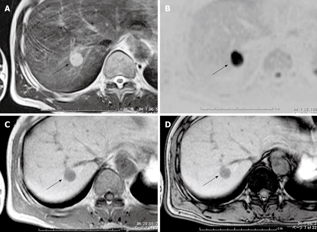
Figure 2 Findings of magnetic resonance imaging (arrows indicate the tumor).
A: The T2-weighted image shows a hyperintense tumor; B: The inverse video of the diffusion-weighted image shows a markedly hyperintense signal; C: In-phase T1-weighted image shows a hypointense signal; D: Opposed-phase T1-weighted image shows no signal intensity reduction compared with Figure 2C.
- Citation: Toriyama E, Nanashima A, Hayashi H, Abe K, Kinoshita N, Yuge S, Nagayasu T, Uetani M, Hayashi T. A case of intrahepatic clear cell cholangiocarcinoma. World J Gastroenterol 2010; 16(20): 2571-2576
- URL: https://www.wjgnet.com/1007-9327/full/v16/i20/2571.htm
- DOI: https://dx.doi.org/10.3748/wjg.v16.i20.2571


