Copyright
©The Author(s) 2017.
World J Gastroenterol. Apr 14, 2017; 23(14): 2493-2504
Published online Apr 14, 2017. doi: 10.3748/wjg.v23.i14.2493
Published online Apr 14, 2017. doi: 10.3748/wjg.v23.i14.2493
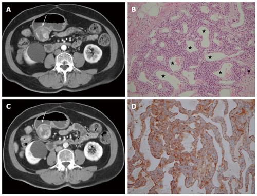
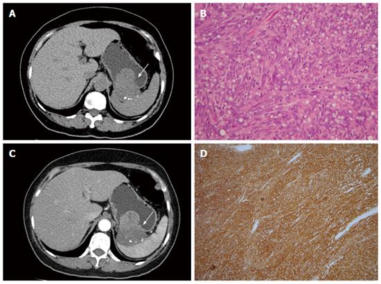
Figure 1 Glomus tumor.
A 66-year-old woman presented with epigastric pain for 1 mo. A: Arterial phase showing a submucosal mass at the gastric antrum (arrow) with an exophytic growth pattern. Peripheral nodular enhancement is evident; B: Portovenous phase showing central fill-in enhancement compared with the arterial phase; C: High power photomicrography (original magnification, × 200, HE stain) showing many vessels (star) filled with red blood cells and lined within the tumor cells. The tumor cells were positive for smooth muscle actin (D).
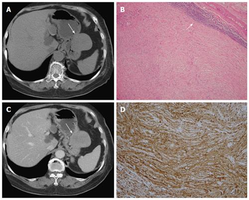
Figure 2 Schwannoma.
A 75-year-old woman presented with coffee ground vomitus. A: Pre-contrast transverse computed tomography (CT) showing a homogeneous iso-density tumor in the greater curvature of the stomach (arrow); B: Post-contrast-enhanced CT showing homogeneously moderate enhancement with a mixed (endoluminal and exophytic) growth pattern; C: Low-power photomicrograph (original magnification, × 20; HE stain) showing that the tumor retains its circumscription with lymphoid aggregate cuffing (arrow); D: The vaguely bundled spindle tumor cells were positive for S-100.
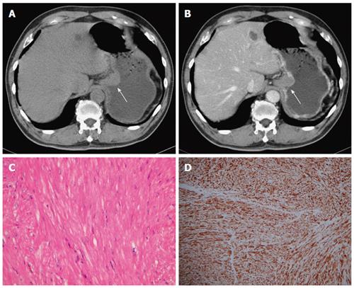
Figure 3 Leiomyoma.
A 70-year-old man had no symptoms. A and B: Pre- and post-contrast-enhanced axial computed tomography scans showing the leiomyoma at the gastric cardia (arrow), with an intraluminal growth pattern and homogeneous, poor enhancement. Note the intact enhancing mucosa, indicating the submucosal lesion; C: High-power photomicrograph (original magnification, × 200; HE stain) showing paucicellular spindle cells with low or moderate cellularity, arranged in perpendicularly oriented fascicles; The tumor cells were positive for smooth muscle actin (D) and negative for CD34 and CD117 (not shown).
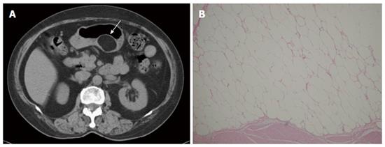
Figure 4 Lipoma.
A 69-year-old man presented with abdominal fullness. A: Non-contrast-enhanced computed tomography (CT) showing a round, sharply marginated, uniform fatty mass (arrow) with negative CT numbers (-90 HU) in the greater curvature of the stomach; B: High-power photomicrograph (original magnification, × 200; HE stain) showing that the tumor consists of mature adipocytes.
Figure 5 Gastrointestinal stromal tumor.
A 58-year-old woman presented with melena and abdominal cramping pain for a year. A: Pre-contrast computed tomography (CT) scan showing amorphous calcifications in a gastric tumor with endoluminal and exophytic growth patterns (arrow); B: Post-contrast-enhanced CT scan showing the intact enhancing mucosa and central necrosis; C: High-power photomicrograph (original magnification, × 100; HE stain) showing spindle cells arranged in lobules; D: The tumor cells were positive for CD117.
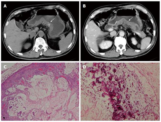
Figure 6 Mucinous adenocarcinoma.
A 65-year-old man presented with vomiting and diarrhea for 2 mo. A and B: Pre- and post-contrast-enhanced computed tomography scans shoings a segmental thickening at the posterior wall of the gastric antrum, with poor enhancement and punctate calcification (arrow). Low-power photomicrograph (original magnification, × 20; HE stain) showing abundant extracellular mucin pools (C) with floating tumor cells and calcifications (D).
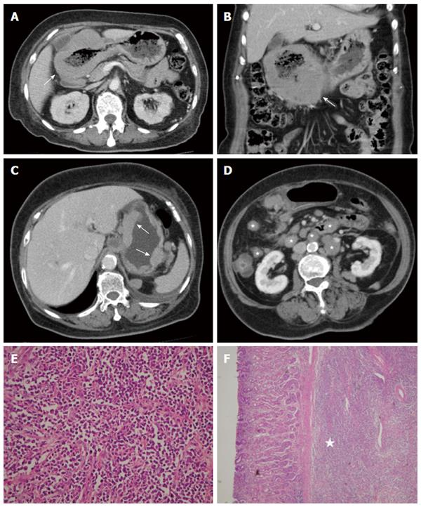
Figure 7 Diffuse large B-cell lymphoma.
A 63-year-old woman presented with epigastralgia. A and B: Contrast-enhanced computed tomography (CT) scan showing diffuse, homogeneous gastric wall thickening with a smooth well-defined outer wall (arrow). An 83-year-old woman presented with tarry stool and constipation for a week; C and D: Post-contrast-enhanced CT revealing wall thickness (arrow) at the gastric body and several enlarged lymph nodes in the mesentery and para-aortic retroperitoneum (stars); E: Low-power photomicrograph (original magnification, × 20; HE stain) showing diffuse proliferation of large monomorphic neoplastic cells with abundant cytoplasms; F: The neoplastic cells occupy the full thickness of the submucosa (star).
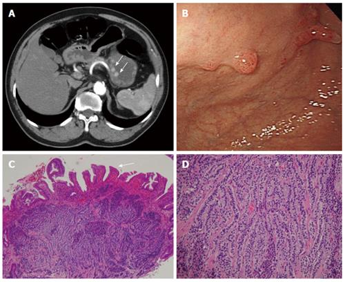
Figure 8 Carcinoid.
A 66-year-old man presented with epigastralgia and elevated levels of serum gastrin. A: Contrast-enhanced transverse and coronal computed tomography (CT) scans showing multiple enhancing polypoid lesions (arrows) at the gastric body; B: Endoscopy showing multiple polypoid lesions; C: Low-power photomicrograph (original magnification, × 10; HE stain) showing atrophic gastritis (atrophy in glandular structures, arrow); D: High-power photomicrograph (original magnification, × 100; HE stain) showing uniform cells bearing round nuclei and growing in a festoon or ribbon-like arrangement in the submucosa.
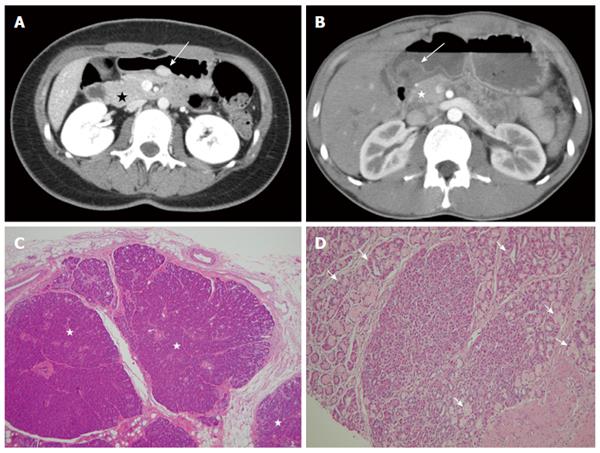
Figure 9 Ectopic pancreas.
A 26-year-old woman presented with postprandial epigastric pain for 2 years. A: Transverse computed tomography (CT) scan showing a small round submucosal lesion with well-defined margins in the wall of the antrum (arrow). Note the contrast material enhancement is higher than that of the normal pancreas (star); B: Low-power photomicrograph (original magnification, × 20; HE stain) showing that pancreas tissue (star) is predominant in the acinar tissue. A 20-year-old man presented with intermittent epigastralgia for 2 mo; C: Transverse CT scan showing a submucosal round mass (arrow) with necrosis at the gastric antrum. Note the poorly enhancing nodular mass, as compared with the markedly enhancing adjacent normal pancreas (star); D: Low-power photomicrograph (original magnification, × 200; HE stain) showing ectopic pancreatic tissue, composed primarily of pancreatic ducts (arrow) in the gastric mucosal layer.
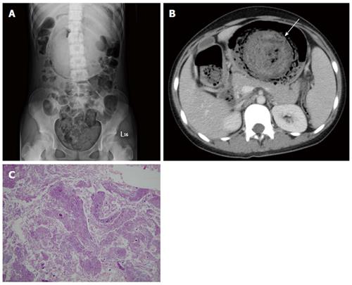
Figure 10 Trichobezoar.
A 13-year-old girl presented with intermittent fever. She exhibited obsessive and compulsive hair pulling. A: Plain film revealing a bezoar outlined by air in the stomach; B: Transverse computed tomography image showing an inhomogeneous mass with a mottled gas pattern in the distended stomach (arrow); C: Low-power photomicrograph (original magnification, × 10; HE stain) showing hair tissue with inflammatory exudate.
- Citation: Lin YM, Chiu NC, Li AFY, Liu CA, Chou YH, Chiou YY. Unusual gastric tumors and tumor-like lesions: Radiological with pathological correlation and literature review. World J Gastroenterol 2017; 23(14): 2493-2504
- URL: https://www.wjgnet.com/1007-9327/full/v23/i14/2493.htm
- DOI: https://dx.doi.org/10.3748/wjg.v23.i14.2493


