Copyright
©The Author(s) 2017.
World J Gastroenterol. Apr 14, 2017; 23(14): 2493-2504
Published online Apr 14, 2017. doi: 10.3748/wjg.v23.i14.2493
Published online Apr 14, 2017. doi: 10.3748/wjg.v23.i14.2493
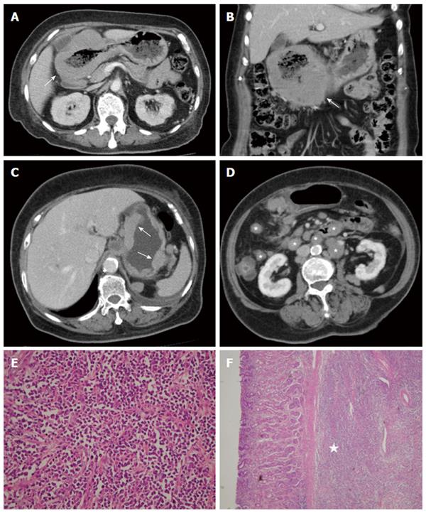
Figure 7 Diffuse large B-cell lymphoma.
A 63-year-old woman presented with epigastralgia. A and B: Contrast-enhanced computed tomography (CT) scan showing diffuse, homogeneous gastric wall thickening with a smooth well-defined outer wall (arrow). An 83-year-old woman presented with tarry stool and constipation for a week; C and D: Post-contrast-enhanced CT revealing wall thickness (arrow) at the gastric body and several enlarged lymph nodes in the mesentery and para-aortic retroperitoneum (stars); E: Low-power photomicrograph (original magnification, × 20; HE stain) showing diffuse proliferation of large monomorphic neoplastic cells with abundant cytoplasms; F: The neoplastic cells occupy the full thickness of the submucosa (star).
- Citation: Lin YM, Chiu NC, Li AFY, Liu CA, Chou YH, Chiou YY. Unusual gastric tumors and tumor-like lesions: Radiological with pathological correlation and literature review. World J Gastroenterol 2017; 23(14): 2493-2504
- URL: https://www.wjgnet.com/1007-9327/full/v23/i14/2493.htm
- DOI: https://dx.doi.org/10.3748/wjg.v23.i14.2493


