
















©The Author(s) 2017.
World J Gastroenterol. Apr 14, 2017; 23(14): 2493-2504
Published online Apr 14, 2017. doi: 10.3748/wjg.v23.i14.2493
Published online Apr 14, 2017. doi: 10.3748/wjg.v23.i14.2493
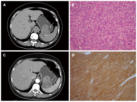
Figure 5 Gastrointestinal stromal tumor.
A 58-year-old woman presented with melena and abdominal cramping pain for a year. A: Pre-contrast computed tomography (CT) scan showing amorphous calcifications in a gastric tumor with endoluminal and exophytic growth patterns (arrow); B: Post-contrast-enhanced CT scan showing the intact enhancing mucosa and central necrosis; C: High-power photomicrograph (original magnification, × 100; HE stain) showing spindle cells arranged in lobules; D: The tumor cells were positive for CD117.
- Citation: Lin YM, Chiu NC, Li AFY, Liu CA, Chou YH, Chiou YY. Unusual gastric tumors and tumor-like lesions: Radiological with pathological correlation and literature review. World J Gastroenterol 2017; 23(14): 2493-2504
- URL: https://www.wjgnet.com/1007-9327/full/v23/i14/2493.htm
- DOI: https://dx.doi.org/10.3748/wjg.v23.i14.2493