Copyright
©2008 The WJG Press and Baishideng.
World J Gastroenterol. Dec 21, 2008; 14(47): 7149-7159
Published online Dec 21, 2008. doi: 10.3748/wjg.14.7149
Published online Dec 21, 2008. doi: 10.3748/wjg.14.7149
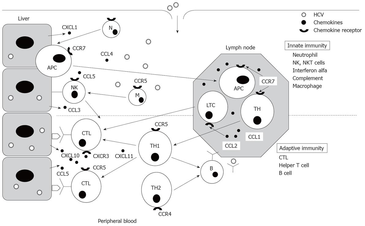
Figure 1 Innate and adaptive immune response.
Importance of specific cytotoxic T cell response. In a non-cytopathic viral infection the development of a vigorous specific cytotoxic T lymphocyte (CTL) response is essential. Professional antigen presenting cells (APC) take up viral antigens and migrate from infected parenchyma to the lymph nodes to prime naive specific CTLs. These cells express the chemokine receptor CCR7 to reach the lymph nodes, attracted by CCL1 and CCL2 chemokines. After priming, specific CTLs lose CCR7 expression and up-regulate CCR5 and CXCR3 chemokine receptors, then migrate to the hepatic parenchyma to develop their effector function, attracted by the chemokines produced in the liver. During the early phase of infection, innate immunity is the first barrier for fighting against the virus. The cellular innate response is also recruited in the infected parenchyma by the interaction between chemokines and their receptors.
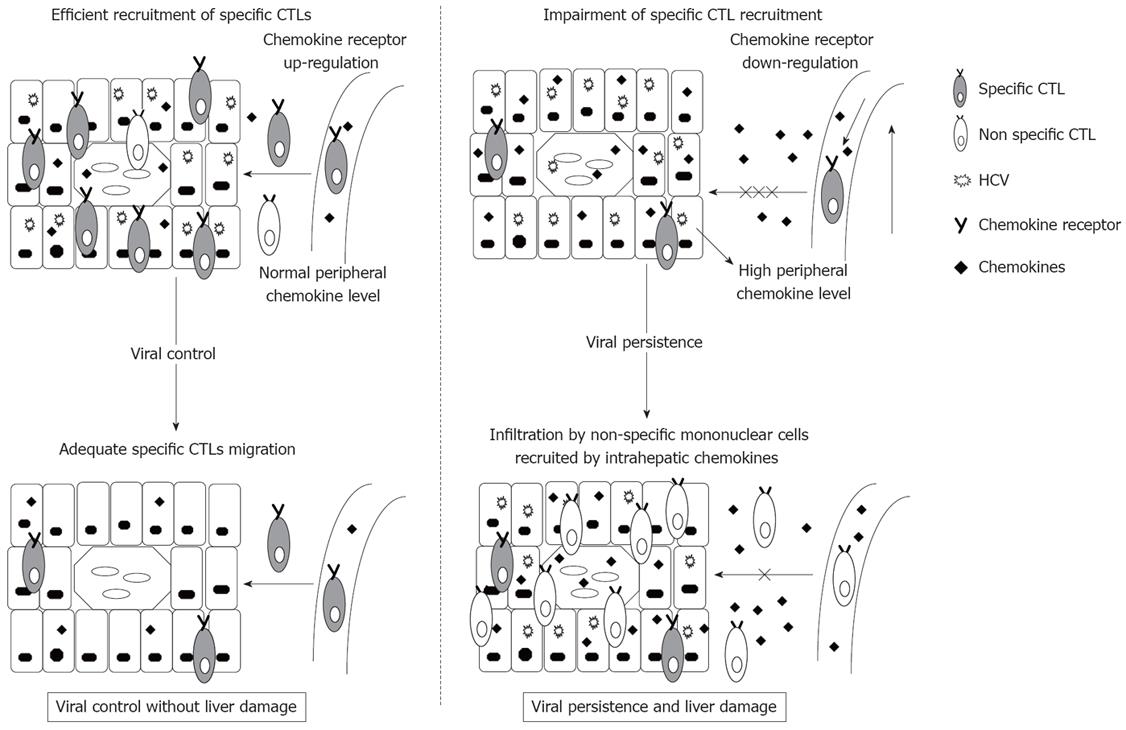
Figure 2 Different migration potential between resolved and persistent HCV infection.
In a resolved infection HCV-specific cytotoxic T lymphocytes (CTL) migrate rapidly to the infected liver, attracted by the chemokines produced by hepatocytes, succeeding in controlling HCV infection without liver damage. In persistent infection the migration of T cells to the liver may be impaired. High chemokine levels during infection may induce chemokine receptor down-regulation on T cells by molecule internalization. This mechanism could impair the migration of specific T cells during primary infection, which could inhibit HCV clearance. During the chronic phase, the persistent presence of HCV in the liver could keep a long-standing chemokine production, which could attract non-specific mononuclear cells to the liver, and be responsible for chronic liver damage. The relative migration impairment due to chemokine receptor down-regulation on non-specific T cells could modulate inflammatory infiltration of the liver, inducing chronic low-grade liver damage, which could favour host and virus long-term survival.
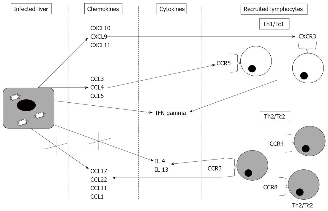
Figure 3 Chemokines and chemokine receptors related to chronic hepatitis C pathogenesis.
In chronic hepatitis C, intrahepatic chemokines recruit Th1/Tc1 cells expressing CCR5/CXCR3 chemokine receptors. The X symbol indicates that chemokines and cytokines associated with a Th2/Tc2 response are not primarily produced in the HCV infected liver.
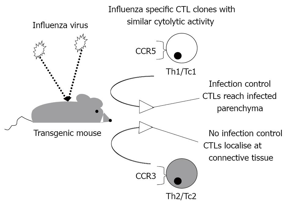
Figure 4 Adequate chemotaxis is necessary to control a viral infection.
Specific cytotoxic T cells with active in vitro effector functionality are not able to control a viral infection if they do not express the appropriate chemokine receptor to reach the infected parenchyma. In a murine model of influenza virus infection, specific CCR5-expressing T cells were able to clear the virus while administration of specific CCR3-expressing T cells induced mouse death. CTLs: cytotoxic T lymphocytes.
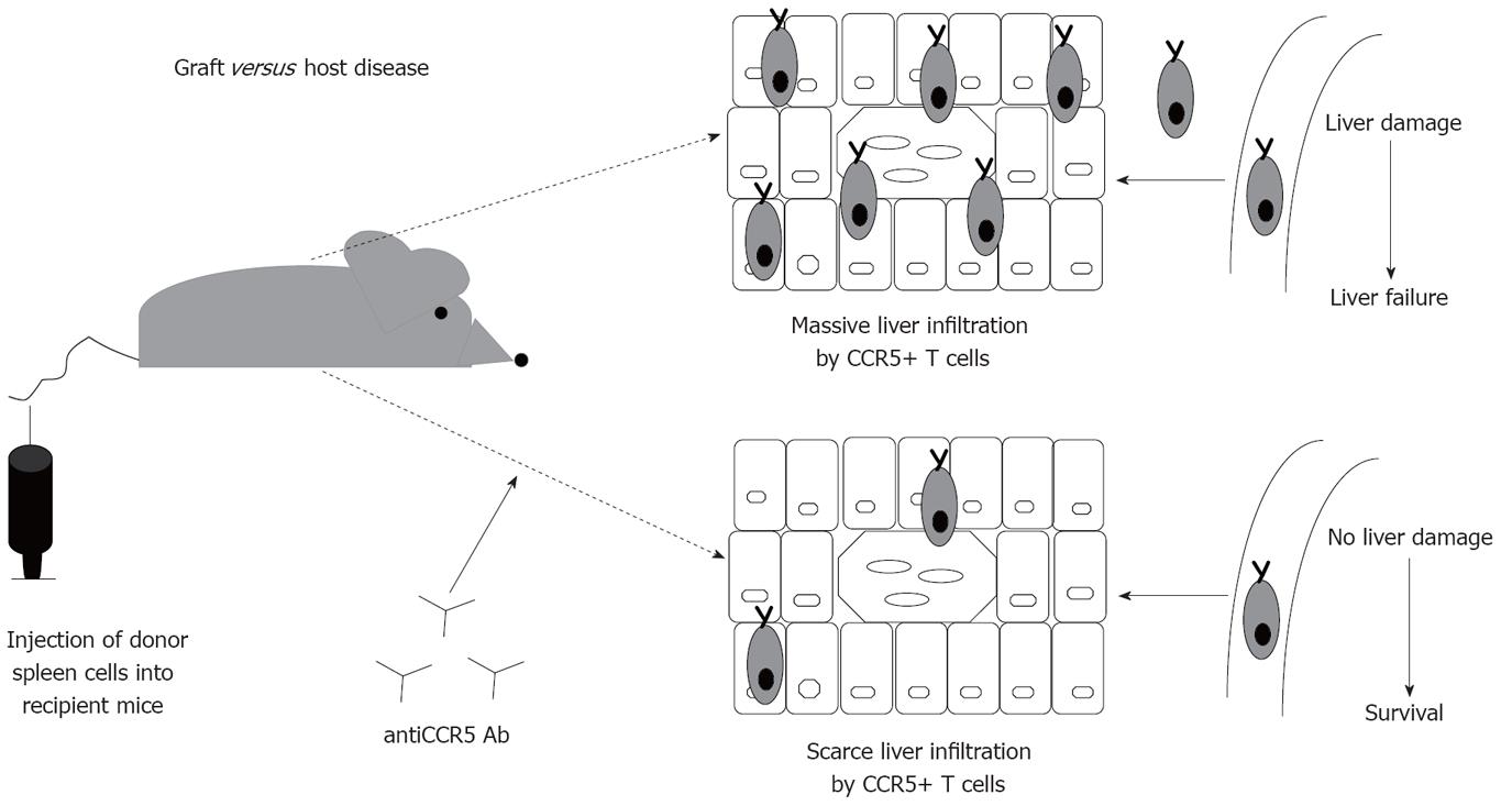
Figure 5 Intrahepatic chemoattraction of non-specific T cells causes liver damage.
In a murine model of graft versus host disease the liver is infiltrated by CCR5-expressing T cells causing liver failure. This process can be blocked by using anti-CCR5 monoclonal antibodies.
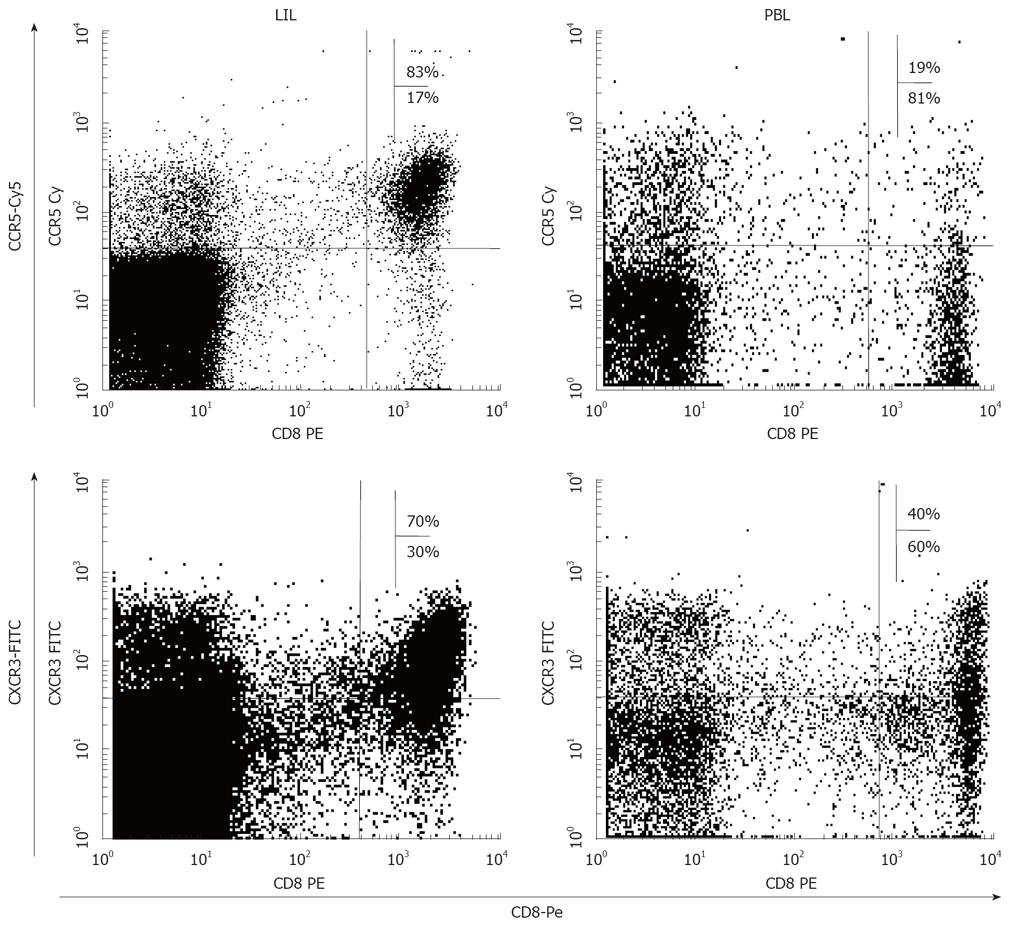
Figure 6 Intrahepatic enrichment of CCR5 and CXCR3 expressing T cells.
FACScan® dot-plots of liver infiltrating (LIL), and peripheral blood (PBL) lymphocytes from a patient with chronic hepatitis C stained with labelled antibodies against CD8, CCR5 and CXCR3. The intrahepatic frequency of CCR5/CXCR3-expressing CD8+ cells is higher than in the peripheral blood.
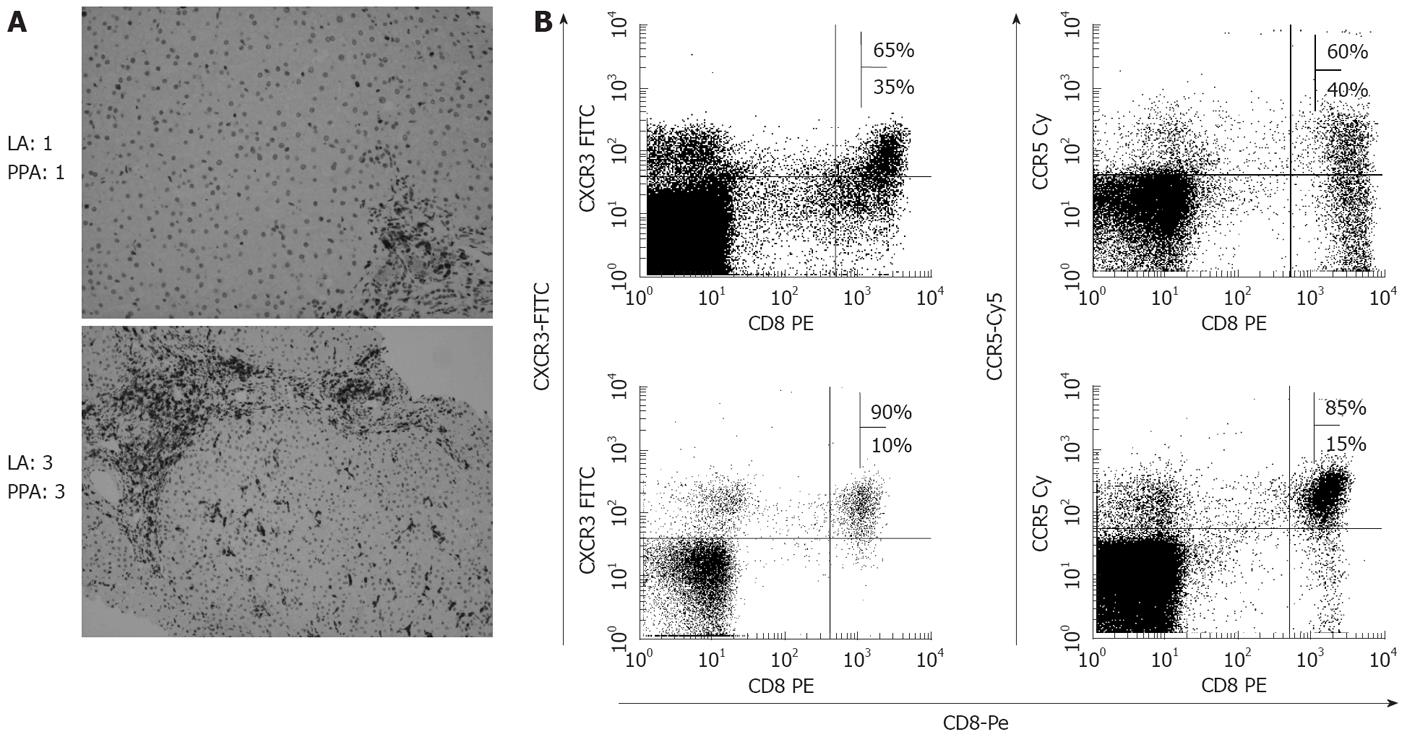
Figure 7 Correlation between liver inflammation and frequency of intrahepatic CCR5 and CXCR3 expressing T cells in chronic hepatitis C.
A: Representative photomicrographs of liver immunostaining for CD8 from two chronic hepatitis C patients with different grades of inflammation. CD8 cells are stained in dark by the immunoperoxidase technique. Original magnification 400X. A patient with lobular activity (LA) 1 and porto-periportal activity (PPA) 1 showed little staining while a patient with LA 3 and PPA 3 presented intense CD8 staining; B: FACScan® dot-plots of intrahepatic lymphocytes from these two patients after staining with CD8-Pe and with either CCR5-Cy5 or CXCR3-FITC mAbs. In the upper right quadrant are represented the double positive cells. The frequency of CCR5/CXCR3 expressing CD8+ cells is higher in the patient with the higher histological liver inflammation.
- Citation: Larrubia JR, Benito-Martínez S, Calvino M, Sanz-de-Villalobos E, Parra-Cid T. Role of chemokines and their receptors in viral persistence and liver damage during chronic hepatitis C virus infection. World J Gastroenterol 2008; 14(47): 7149-7159
- URL: https://www.wjgnet.com/1007-9327/full/v14/i47/7149.htm
- DOI: https://dx.doi.org/10.3748/wjg.14.7149


