Copyright
©The Author(s) 2004.
World J Gastroenterol. Mar 15, 2004; 10(6): 825-829
Published online Mar 15, 2004. doi: 10.3748/wjg.v10.i6.825
Published online Mar 15, 2004. doi: 10.3748/wjg.v10.i6.825
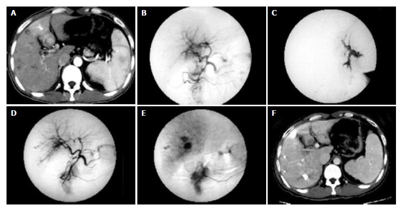
Figure 1 Multiple HCC nodules in a 34-year-old patient.
A: CT image obtained during the arterial phase showed the predominant enhancement of the medial segment of left lobe and the enhancement of left portal branches, which represent APS. B: Hepatic arteriogram demonstrated the arterioportal shunt. C: The microcatheter was inserted into the feeding artery of APS. DSA showed the strong or fast blood flow of APS. D-E: Hepatic arteriogram showed that the arterioportal shunt was no longer visible after embolization with a microcoil and ethanol. F: Follow-up CT scan showed lipiodol accumulation in multiple HCC nodules, and liver necrosis was not seen in the distribution of the hepatic artery which had been treated with ethanol.
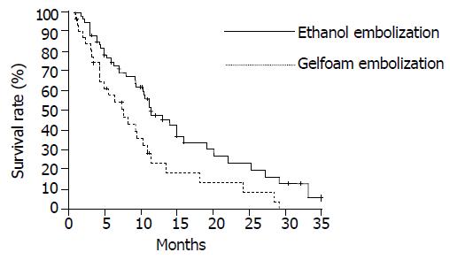
Figure 2 Cumulative survival curves for patients with HCC and APS in two therapeutic groups are shown.
The survival rates for patients in the ethanol group were significantly higher than those in the Gelfoam group (P < 0.05).
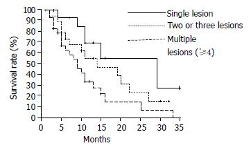
Figure 3 Cumulative survival curves for patients with HCC and APS in relation to number of tumors are shown in ethanol group.
The survival rates of patients with monofocal HCC and APS were significantly higher than those of patients with multifocal HCC and APS (single lesion vs two or three lesions, P < 0.05; two or three lesions vs four or more lesions, P < 0.05).
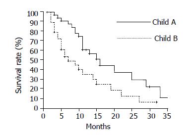
Figure 4 Cumulative survival curves for patients with HCC and APS according to the Child class were shown in ethanol group.
The survival rates of patients of Child class A were higher than those of Child class B (P < 0.05).
- Citation: Huang MS, Lin Q, Jiang ZB, Zhu KS, Guan SH, Li ZR, Shan H. Comparison of long-term effects between intra-arterially delivered ethanol and Gelfoam for the treatment of severe arterioportal shunt in patients with hepatocellular carcinoma. World J Gastroenterol 2004; 10(6): 825-829
- URL: https://www.wjgnet.com/1007-9327/full/v10/i6/825.htm
- DOI: https://dx.doi.org/10.3748/wjg.v10.i6.825


