
















©The Author(s) 2004.
World J Gastroenterol. Mar 15, 2004; 10(6): 825-829
Published online Mar 15, 2004. doi: 10.3748/wjg.v10.i6.825
Published online Mar 15, 2004. doi: 10.3748/wjg.v10.i6.825
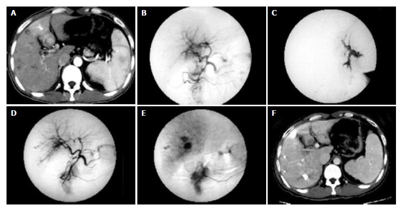
Figure 1 Multiple HCC nodules in a 34-year-old patient.
A: CT image obtained during the arterial phase showed the predominant enhancement of the medial segment of left lobe and the enhancement of left portal branches, which represent APS. B: Hepatic arteriogram demonstrated the arterioportal shunt. C: The microcatheter was inserted into the feeding artery of APS. DSA showed the strong or fast blood flow of APS. D-E: Hepatic arteriogram showed that the arterioportal shunt was no longer visible after embolization with a microcoil and ethanol. F: Follow-up CT scan showed lipiodol accumulation in multiple HCC nodules, and liver necrosis was not seen in the distribution of the hepatic artery which had been treated with ethanol.
- Citation: Huang MS, Lin Q, Jiang ZB, Zhu KS, Guan SH, Li ZR, Shan H. Comparison of long-term effects between intra-arterially delivered ethanol and Gelfoam for the treatment of severe arterioportal shunt in patients with hepatocellular carcinoma. World J Gastroenterol 2004; 10(6): 825-829
- URL: https://www.wjgnet.com/1007-9327/full/v10/i6/825.htm
- DOI: https://dx.doi.org/10.3748/wjg.v10.i6.825