Copyright
©The Author(s) 2004.
World J Gastroenterol. Feb 1, 2004; 10(3): 361-365
Published online Feb 1, 2004. doi: 10.3748/wjg.v10.i3.361
Published online Feb 1, 2004. doi: 10.3748/wjg.v10.i3.361
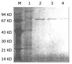
Figure 1 SDS-PAGE analysis of HSP70-tumor peptide com-plexes (silver staining).
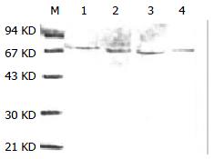
M: protein molecular weight marker, 1: protein eluted with 250-350 mmol/L NaCl buffer, after ADP-agarose chromatography (eluted with 0.5 mol/L NaCl buffer, PH7.5, containing 20 mmol/L Tris-acetate) and DEAE-ion exchange, 2-4: protein eluted with 250-350 mmol/L NaCl buffer, after ADP-agarose chromatography (eluted with 3 mmol ADP/L buffer, pH7.5, containing 20 mmol/L Tris-acetate) and DEAE-ion exchange.
Figure 2 Western blot identification of HSP70-tumor peptide complexes purified from HCaF cells.
The notes are the same with Figure 1.
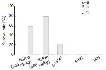
Figure 3 Immunoprotective effect of HSP70-associated pep-tides against tumor.
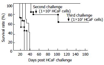
Figure 4 Protective effect of transferred immune spleen cells against repeated challenges with HCaF.
—▲―▲ ×_× First, second, and third challenge controls. ●_● Transferred immune spleen cell group.
- Citation: Chen DX, Su YR, Shao GZ, Qian ZC. Purification of heat shock protein 70-associated tumor peptides and its antitumor immunity on hepatoma in mice. World J Gastroenterol 2004; 10(3): 361-365
- URL: https://www.wjgnet.com/1007-9327/full/v10/i3/361.htm
- DOI: https://dx.doi.org/10.3748/wjg.v10.i3.361


