Copyright
©The Author(s) 2004.
World J Gastroenterol. Jan 15, 2004; 10(2): 299-302
Published online Jan 15, 2004. doi: 10.3748/wjg.v10.i2.299
Published online Jan 15, 2004. doi: 10.3748/wjg.v10.i2.299
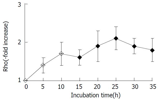
Figure 1 Changes in levels of Rho family protein in SMMC-7721cells induced by LPA.
The levels of Rho protein were ana-lyzed by Western blot analysis. The data shown are expressed as -x±s from three different experiments.
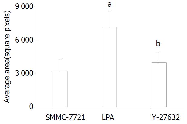
Figure 2 Tumor cell random motility determined by gold-col-loid assay.
Cells were seeded onto the coverslips and allowed to adhere for 1 h and then incubated in FBS containing LPA with or without Y-27632 for 24 h. The areas of clearing in the gold colloid corresponding to phagokinetic cell tracks were counted and represented as -x±s from triplicate experiments. aP < 0.01 LPA treated cells vs SMMC-7721 control cells, bP < 0.01 Y-27632 treated cells vs LPA treated cells.
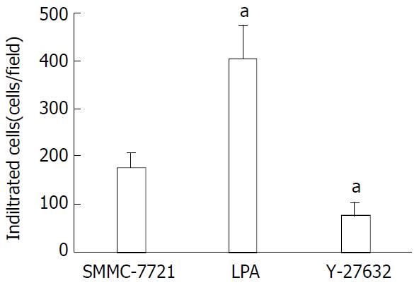
Figure 3 Inhibition of LPA-induced tumor cell invasion by Y-27632.
Serum-starved cells were seeded onto porous filters. After incubation of the filters with Y-27632 in the presence of LPA 25 μM for 4.5 h to permit penetration of the cells, nonmigrating cells were removed from the upper chamber and migrating cells adherent to the underside of the filters were counted in a minimum of 10-high power fields. Data were expressed as relative migration (number of cells/field) and represented as -x±s from triplicate experiments. aP < 0.01 relative to SMMC-7721 control cells.
- Citation: Wang DS, Dou KF, Li KZ, Song ZS. Enhancement of migration and invasion of hepatoma cells via a Rho GTPase signaling pathway. World J Gastroenterol 2004; 10(2): 299-302
- URL: https://www.wjgnet.com/1007-9327/full/v10/i2/299.htm
- DOI: https://dx.doi.org/10.3748/wjg.v10.i2.299


