
















Copyright: ©Author(s) 2026.
Artif Intell Cancer. Sep 8, 2026; 7(1): 122429
Published online Sep 8, 2026. doi: 10.35713/aic.122429
Published online Sep 8, 2026. doi: 10.35713/aic.122429
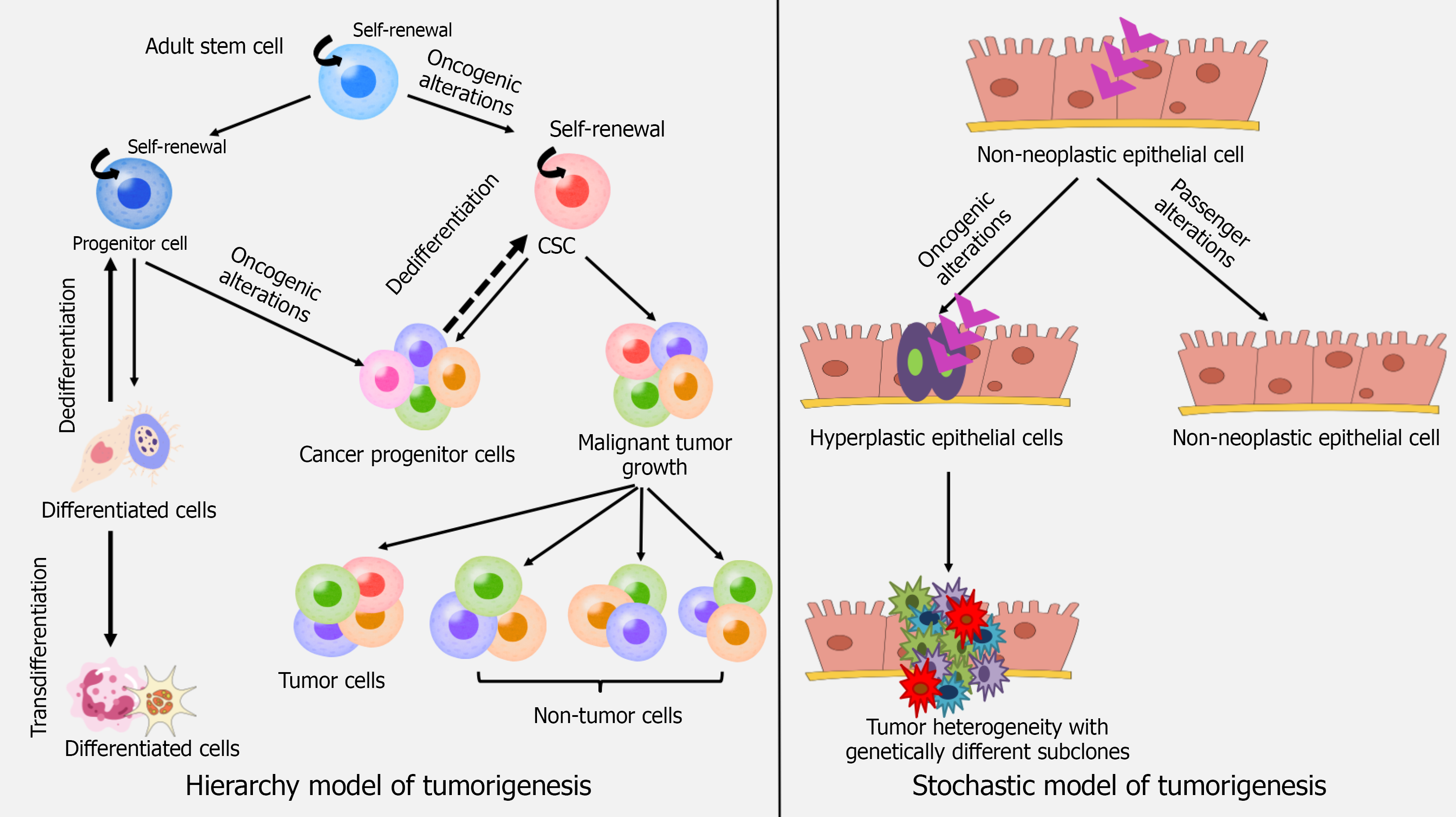
Figure 1 Schematic representation of hierarchy model and stochastic model of tumorigenesis.
This figure was prepared on power point and Canva. Two foundational models of tumorigenesis explain the origin and growth of tumors as well as maintenance of cellular diversity. Hierarchy model proposes the ability of only specialized subpopulation of cells to drive the tumor development. The stochastic model argues that any cell has an equal and random chance to promote tumor development. CSC: Cancer stem cell.
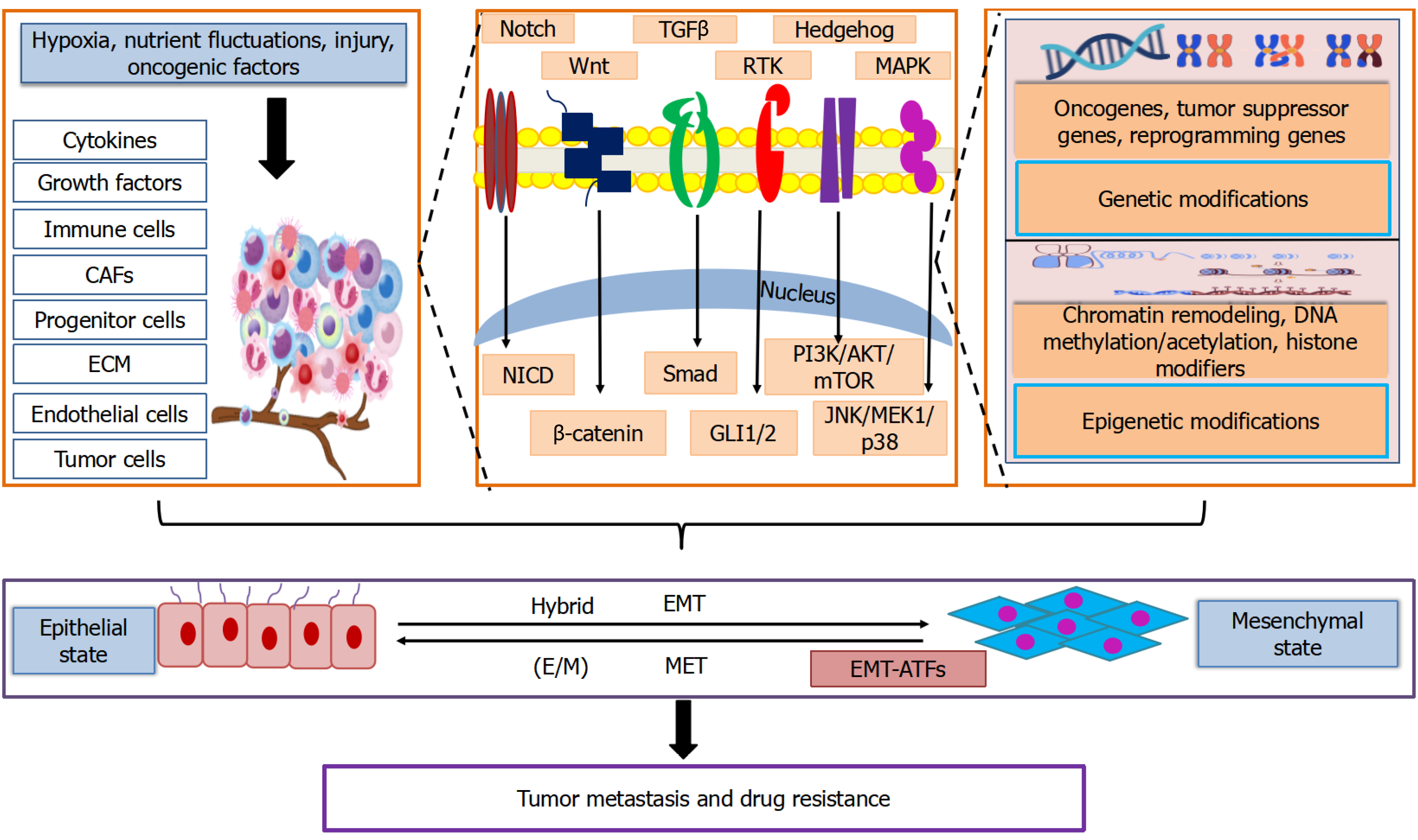
Figure 2 Mechanistic insights of the processes which regulate cancer cell plasticity in tumor microenvironment.
This figure was prepared on power point and Canva. Environmental cues in a tumor ecosystem such as hypoxia and nutrients fluctuations or injury and oncogenic insults activate signaling pathways such as Notch, Wnt, mitogen-activated protein kinase, receptor tyrosine kinase, hedgehog and transforming growth factor beta. Dynamic tumor microenvironment results in changes in genetic, epigenetic, transcriptional and translational landscape enabling the cells to undergo epithelial-mesenchymal transition, mesenchymal-epithelial transition and partial/hybrid transition to promote cancer cell plasticity, invasion and metastatic potential and therapy resistance. AKT: Protein kinase B; CAFs: Cancer associated fibroblasts; ECM: Extracellular matrix; E/M: Partial/hybrid; EMT: Epithelial-mesenchymal transition; EMT-ATFs: Epithelial-to-mesenchymal transition-associated transcription factors; GLI1/2: Glioma-associated oncogene homolog 1 and glioma-associated oncogene homolog 2; JNK: Jun N-terminal kinase; MAPK: Mitogen-activated protein kinase; MEK1: Mitogen-activated protein kinase kinase 1; MET: Mesenchymal-epithelial transition; mTOR: Mammalian target of rapamycin, NICD: Notch intracellular domain; PI3K: Phosphoinositide 3-kinase; RTK: Receptor tyrosine kinase; Smad: Suppressor of mother against decapentaplegic; TGFβ: Transforming growth factor beta.
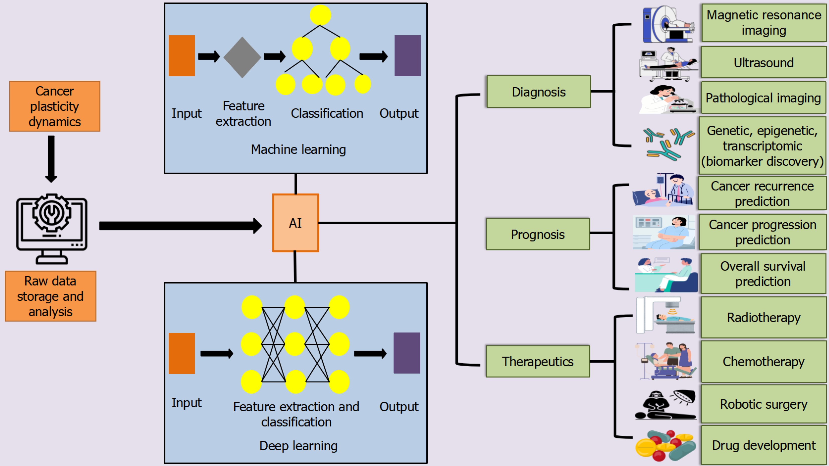
Figure 3 Artificial intelligence and its applications in cancer diagnostics, prognostics and therapeutics.
This figure was prepared on power point and Canva. Machine learning and Deep learning models are two important subsets of artificial intelligence that enable the computers to learn and interpret the experimental data on cancer cell plasticity dynamics to provide tailored diagnosis, risk assessment, accelerating drug discovery and individualizing treatment regimens. AI: Artificial intelligence.
- Citation: Srivastava M, Garg M. Artificial intelligence-driven decoding of trajectories of cancer cell plasticity: A paradigm shift in oncological assessment and treatment. Artif Intell Cancer 2026; 7(1): 122429
- URL: https://www.wjgnet.com/2644-3228/full/v7/i1/122429.htm
- DOI: https://dx.doi.org/10.35713/aic.122429