Copyright
©The Author(s) 2022.
Artif Intell Cancer. Jul 28, 2022; 3(3): 42-53
Published online Jul 28, 2022. doi: 10.35713/aic.v3.i3.42
Published online Jul 28, 2022. doi: 10.35713/aic.v3.i3.42
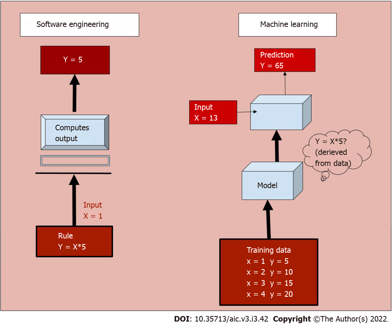
Figure 1 Functioning of a software engineering process vs a machine learning model.
In the former, a rule is programmed into the system based on which it computes output for a given input. Whereas in a machine learning model, the system learns from a large training dataset, and deciphers the rule to predict output.
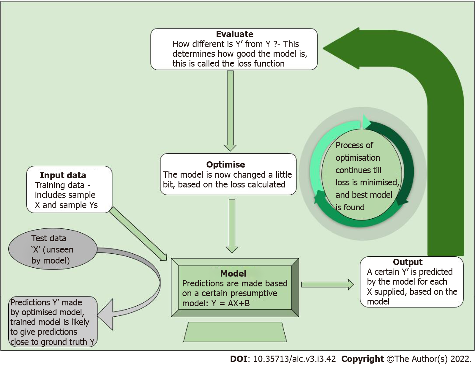
Figure 2 Process of training a machine learning model.
Following the initial training dataset, the model is tested on unseen data. The predicted output is compared to desired output, and changes are incorporated into the model till the predicted output is sufficiently close to desired output.
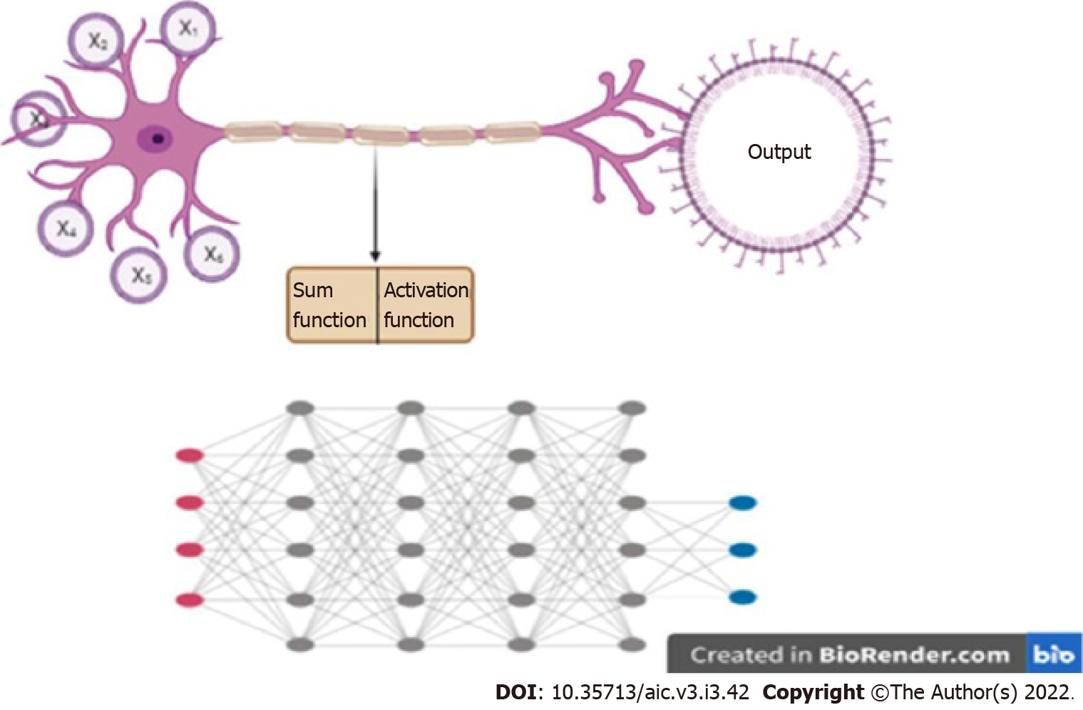
Figure 3 Deep learning.
A: Artificial neural networks take input from a number of channels (x1....xn), perform a mathematical function, and decide on ‘firing’ based on activation function; B: Deep learning involves multiple layers of such neural networks with many nodes. Each node communicates with all nodes of the connecting networks. Created with BioRender.com.
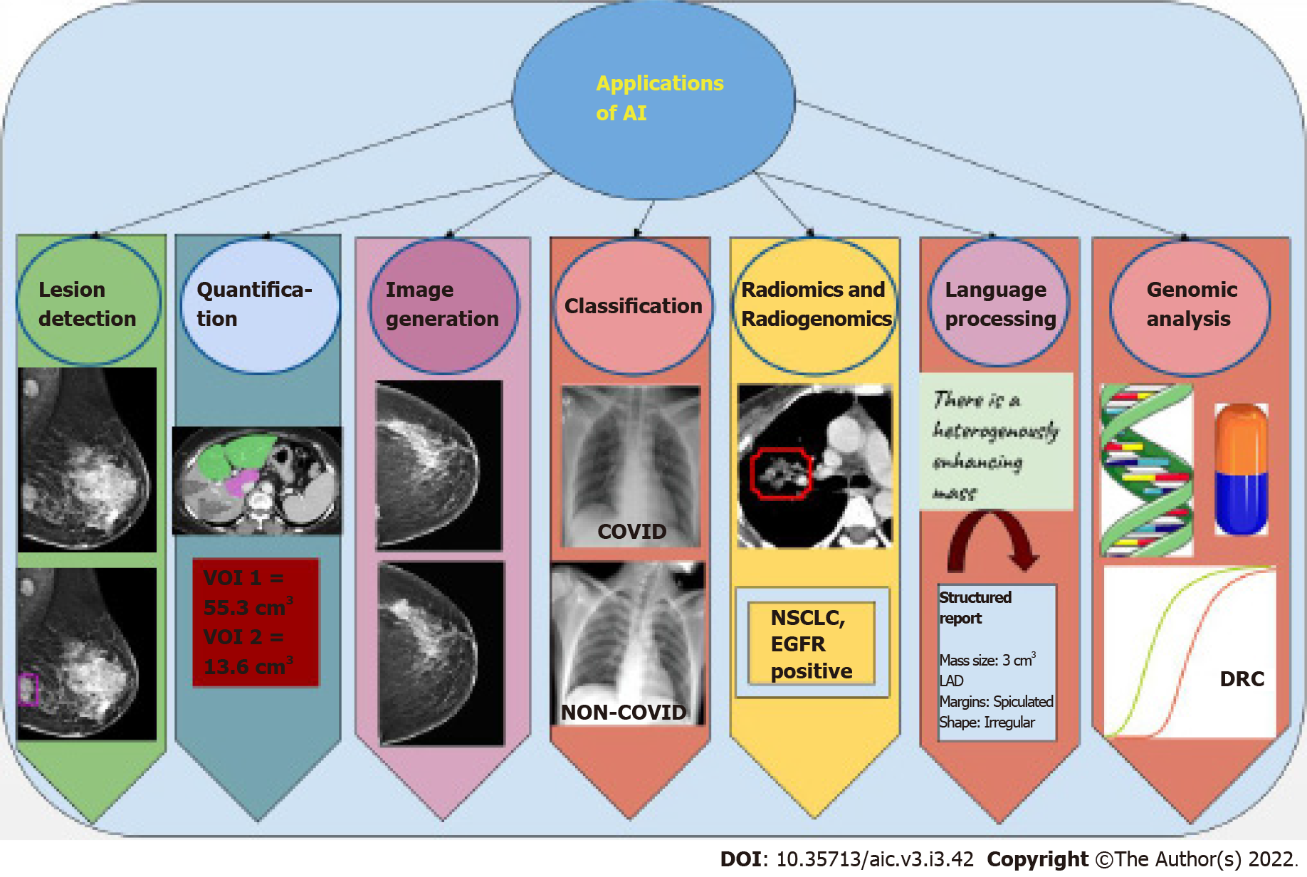
Figure 4 Summary of tasks performed by artificial intelligence networks.
The applications are diverse and range from lesion detection, i.e., automatic identification of the area of pathology in a particular image, to segmentation of defined areas and quantification of area, volume, or percentage segment involved. Synthetic images may also be generated by artificial intelligence (AI) networks that closely resemble natural images. Further, it performs classification tasks, wherein images are placed into one of two or three categories. Analysis of texture features and correlation with genetic mutations is also possible using AI. Newer applications include language processing, from free flow to structured reports with standard, reproducible terminology. Genomic analysis of large amounts of DNA to determine drug response and susceptibility to drugs is also an evolving application. AI: Artificial intelligence; VOI: Volume of interest; NSCLC: Non-small cell lung carcinoma; EGFR: Epidermal growth factor receptor; LAD: Long axis diameter; DRC: Dose response curve.
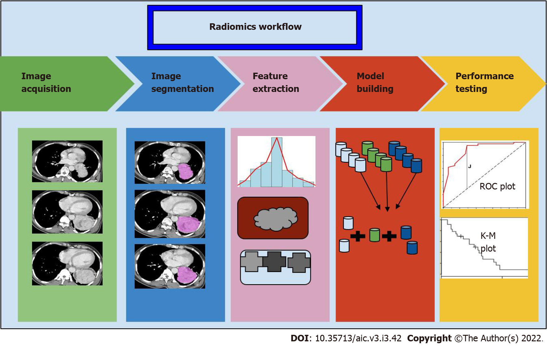
Figure 5 The process of radiomics begins with segmentation of the region of interest from the image volumes, which may be manual or automatic.
Following this, a series of features both histogram based and pixel based are extracted, and a set of these are chosen as classifiers for the discriminator model. The performance of this model is tested on a different set of data using statistical methods. ROC: Receiver operating characteristic; K-M: Kaplan-Meier
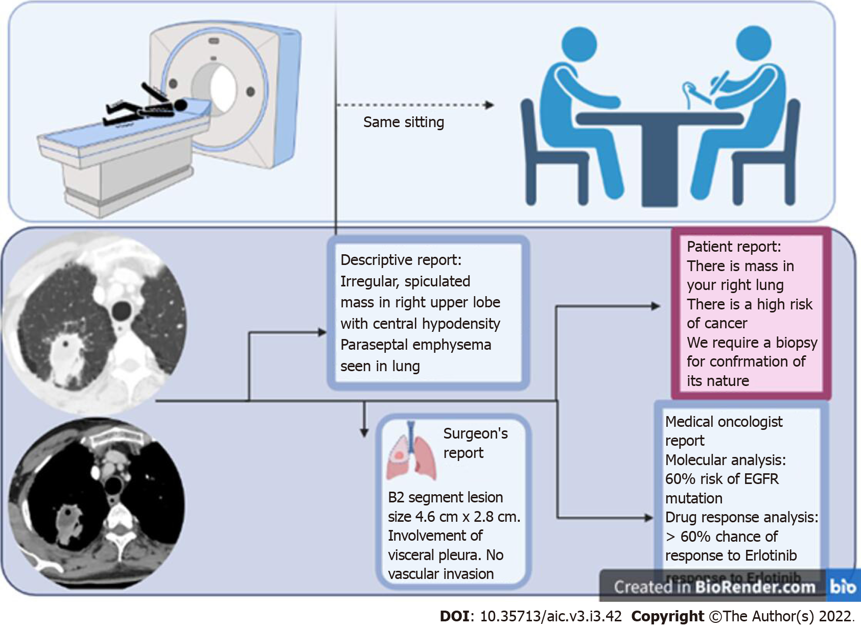
Figure 6 Artificial intelligence enables the transition into the era of personalised medicine, where the assistance of artificial intelligence allows a radiologist to interact with the patient, correlate the findings with the clinical setting, and potentially issue a report in the same sitting as an imaging scan.
Multiple different formats of report may be created based on the target audience, which enable clear communication and enhanced information exchange.
- Citation: Ramachandran A, Bhalla D, Rangarajan K, Pramanik R, Banerjee S, Arora C. Building and evaluating an artificial intelligence algorithm: A practical guide for practicing oncologists. Artif Intell Cancer 2022; 3(3): 42-53
- URL: https://www.wjgnet.com/2644-3228/full/v3/i3/42.htm
- DOI: https://dx.doi.org/10.35713/aic.v3.i3.42


