Copyright
©The Author(s) 2019.
World J Clin Cases. Jul 6, 2019; 7(13): 1652-1659
Published online Jul 6, 2019. doi: 10.12998/wjcc.v7.i13.1652
Published online Jul 6, 2019. doi: 10.12998/wjcc.v7.i13.1652
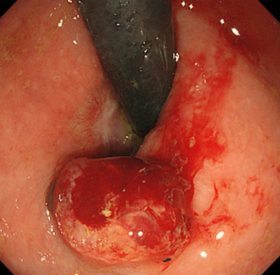
Figure 7 Follow-up endoscopic findings 6 mo after the endoscopic submucosal dissection.
A 10-mm protruded lesion is identified about 10 mm from the oral side of the post-endoscopic submucosal dissection scar. Biopsy specimens from this lesion confirm a malignant melanoma.
- Citation: Manabe S, Boku Y, Takeda M, Usui F, Hirata I, Takahashi S. Endoscopic submucosal dissection as excisional biopsy for anorectal malignant melanoma: A case report. World J Clin Cases 2019; 7(13): 1652-1659
- URL: https://www.wjgnet.com/2307-8960/full/v7/i13/1652.htm
- DOI: https://dx.doi.org/10.12998/wjcc.v7.i13.1652


