Copyright
©The Author(s) 2024.
World J Clin Cases. Oct 26, 2024; 12(30): 6374-6382
Published online Oct 26, 2024. doi: 10.12998/wjcc.v12.i30.6374
Published online Oct 26, 2024. doi: 10.12998/wjcc.v12.i30.6374
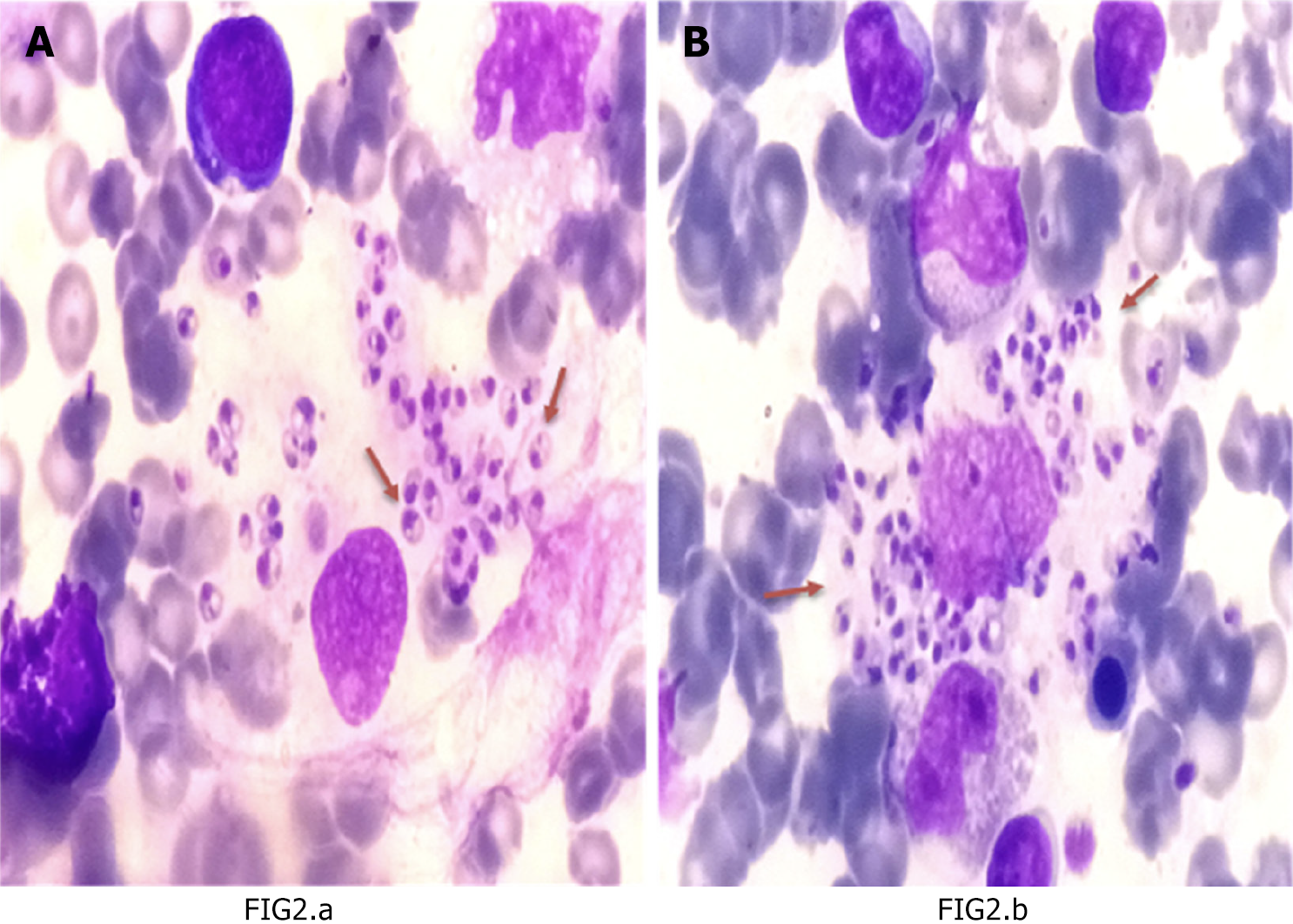
Figure 2 Bone marrow smears (Giemsa stain × 100).
A: Reputed macrophage shedding Extracellular amastigotes, each with a prominent nucleus and kinetoplast (red arrows); B: Leishmania donovani bodies as intracytoplasmic, non-flagellated leishmania parasites (amastigotes), located intracellularly in macrophages (red arrows).
- Citation: Elnoor ZIA, Abdelmajeed O, Mustafa A, Gasim T, Musa SAM, Abdelmoneim AH, Omer IIA, Fadl HAO. Hematological picture of pediatric Sudanese patients with visceral leishmaniasis and prediction of leishmania donovani parasite load. World J Clin Cases 2024; 12(30): 6374-6382
- URL: https://www.wjgnet.com/2307-8960/full/v12/i30/6374.htm
- DOI: https://dx.doi.org/10.12998/wjcc.v12.i30.6374


