Copyright
©The Author(s) 2022.
World J Clin Cases. Sep 6, 2022; 10(25): 9012-9019
Published online Sep 6, 2022. doi: 10.12998/wjcc.v10.i25.9012
Published online Sep 6, 2022. doi: 10.12998/wjcc.v10.i25.9012
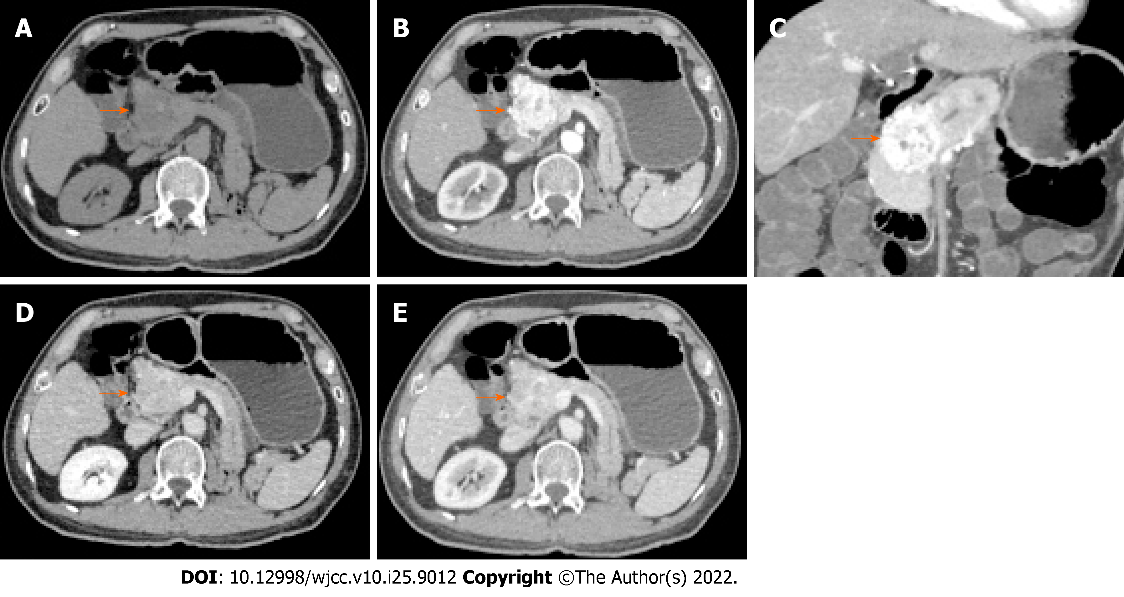
Figure 2 Abdominal computed tomography.
A: Plain scan; B: Arterial phase; C: Coronal plane; D: Venous phase; and E: Delayed phase. A low-density mass is seen (arrow). There is marked heterogeneous enhancement in the “fast in and fast out” mode; multiple tortuous blood vessels are present at the periphery and small patchy non-enhancing areas in the middle (indicating necrosis). There is moderately decreased enhancement in the venous and delayed phases.
- Citation: Liang XK, Li LJ, He YM, Xu ZF. Misdiagnosis of pancreatic metastasis from renal cell carcinoma: A case report. World J Clin Cases 2022; 10(25): 9012-9019
- URL: https://www.wjgnet.com/2307-8960/full/v10/i25/9012.htm
- DOI: https://dx.doi.org/10.12998/wjcc.v10.i25.9012


