Copyright
©The Author(s) 2022.
World J Clin Cases. Apr 16, 2022; 10(11): 3630-3638
Published online Apr 16, 2022. doi: 10.12998/wjcc.v10.i11.3630
Published online Apr 16, 2022. doi: 10.12998/wjcc.v10.i11.3630
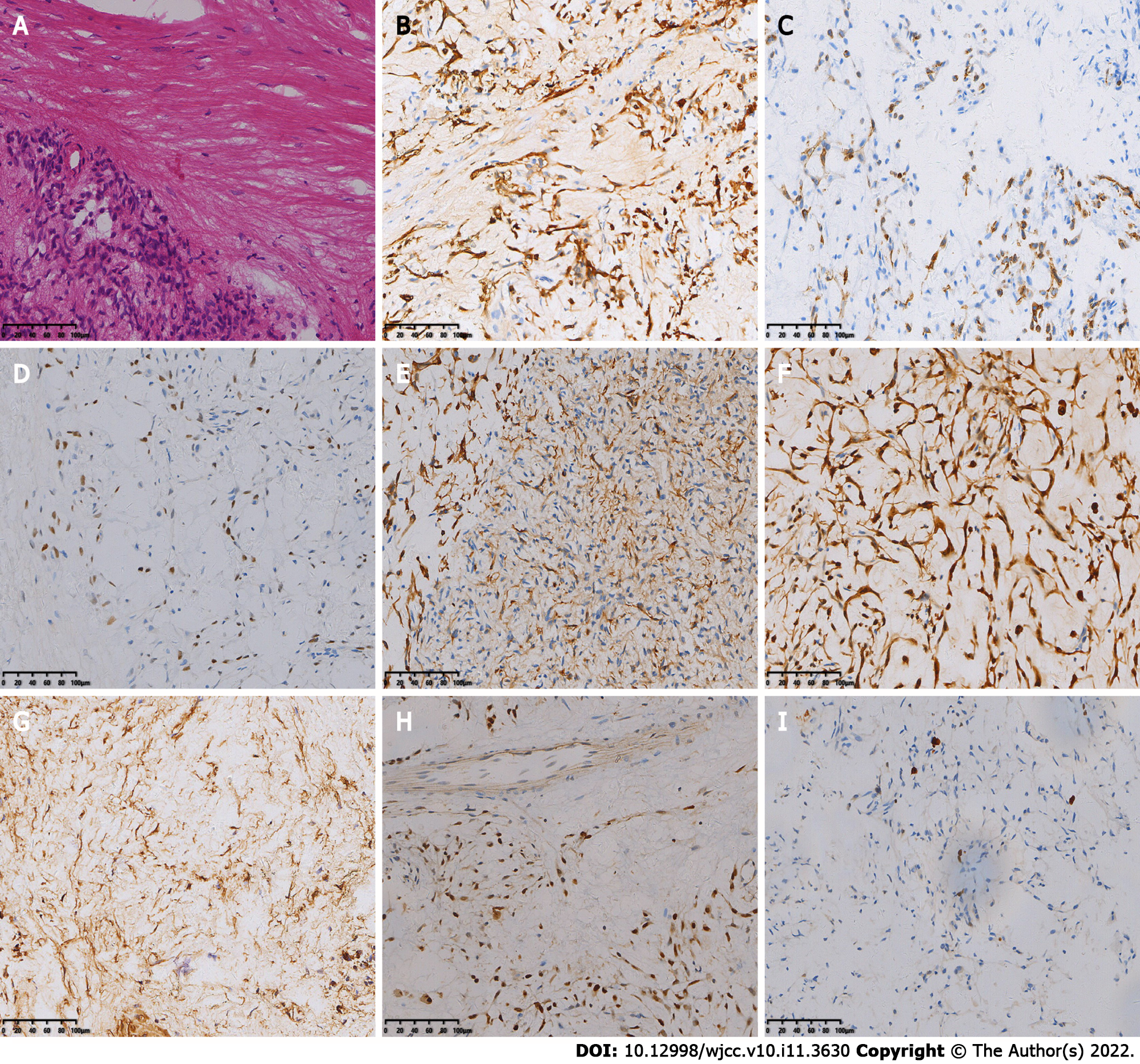
Figure 2 The first postoperative immunohistochemistry results.
A: Hematoxylin-eosin staining shows spindle-shaped tumor cells, mucinous mesenchyme and foci of necrosis, with nuclear schizophrenia and marked nuclear anisotropy, and a small localized pleomorphic adenomatous component in the tumor (magnification × 200); B-I: Immunohistochemistry showed that the tumor cells were positive for CK (B), CK7 (C), P63 (D), Calponin (E), S-100 (F), SMA (G), SOX-10 (H) and 5% of cells were positive for Ki-67 (I) [Envision (B-I) × 200].
- Citation: Huang WP, Li LM, Gao JB. Pleomorphic adenoma of the left lacrimal gland recurred and transformed into myoepithelial carcinoma after multiple operations: A case report. World J Clin Cases 2022; 10(11): 3630-3638
- URL: https://www.wjgnet.com/2307-8960/full/v10/i11/3630.htm
- DOI: https://dx.doi.org/10.12998/wjcc.v10.i11.3630


