Copyright
©The Author(s) 2021.
World J Clin Cases. Mar 16, 2021; 9(8): 1916-1922
Published online Mar 16, 2021. doi: 10.12998/wjcc.v9.i8.1916
Published online Mar 16, 2021. doi: 10.12998/wjcc.v9.i8.1916
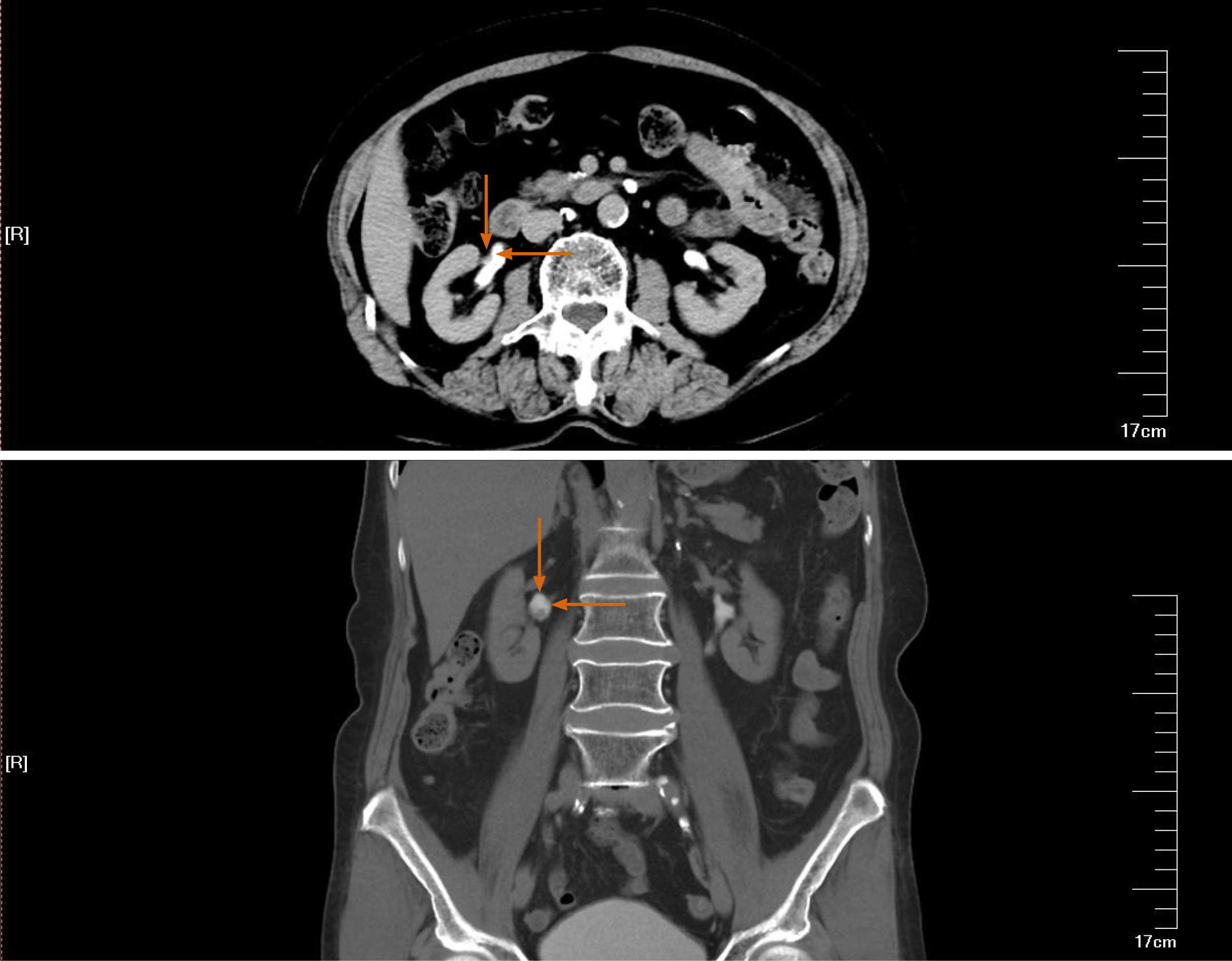
Figure 1 Computed tomography scan of the excretory urography stage.
The arrow indicates a right renal pelvic tumor.
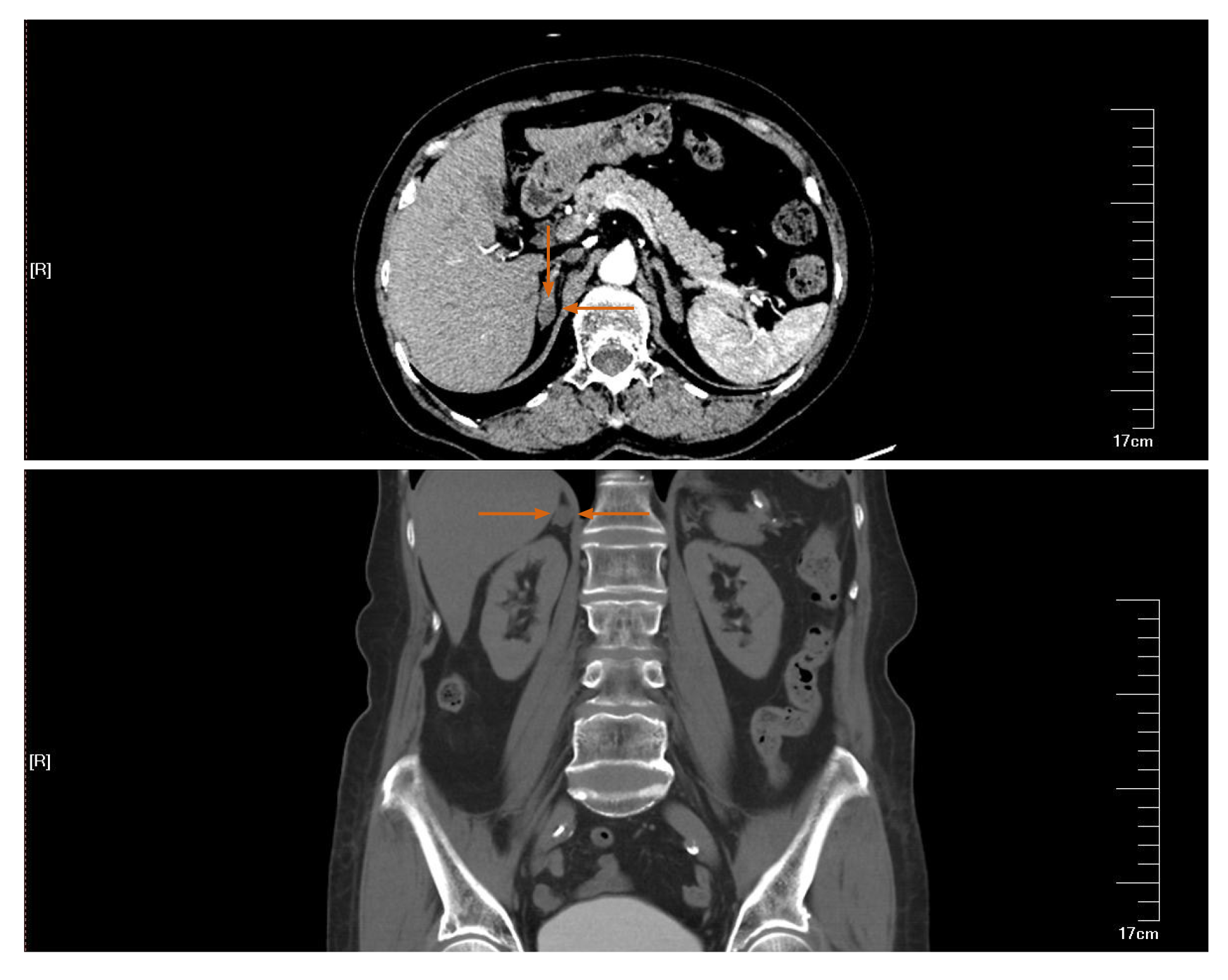
Figure 2 Computed tomography scan in the artery phase.
The arrow indicates a right adrenal gland adenoma.
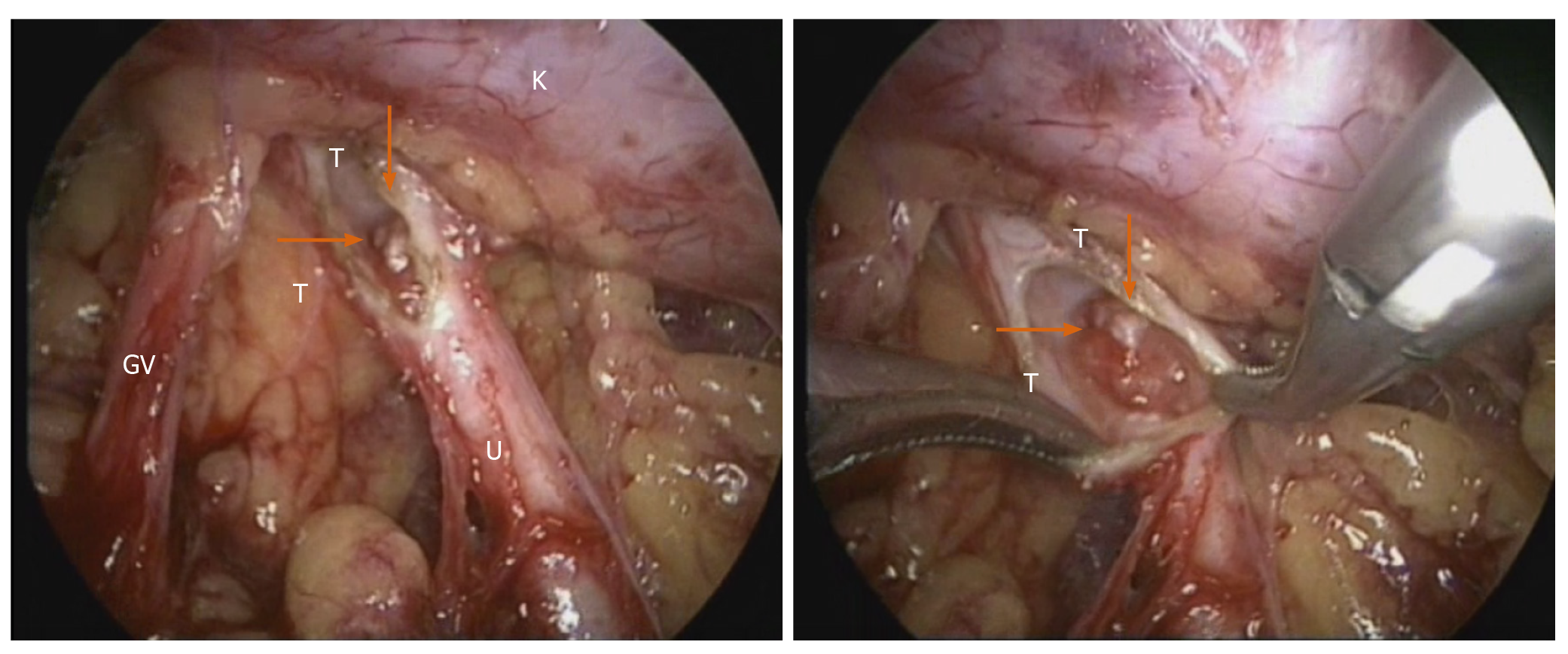
Figure 3 Intraoperative view of the right retroperitoneal space.
GV: Genital vein; K: Kidney; T: Tumor; U: Ureter.
- Citation: Wang YL, Zhang HL, Du H, Wang W, Gao HF, Yu GH, Ren Y. Retroperitoneal laparoscopic partial resection of the renal pelvis for urothelial carcinoma: A case report. World J Clin Cases 2021; 9(8): 1916-1922
- URL: https://www.wjgnet.com/2307-8960/full/v9/i8/1916.htm
- DOI: https://dx.doi.org/10.12998/wjcc.v9.i8.1916


