Copyright
©The Author(s) 2021.
World J Clin Cases. Oct 26, 2021; 9(30): 9198-9204
Published online Oct 26, 2021. doi: 10.12998/wjcc.v9.i30.9198
Published online Oct 26, 2021. doi: 10.12998/wjcc.v9.i30.9198
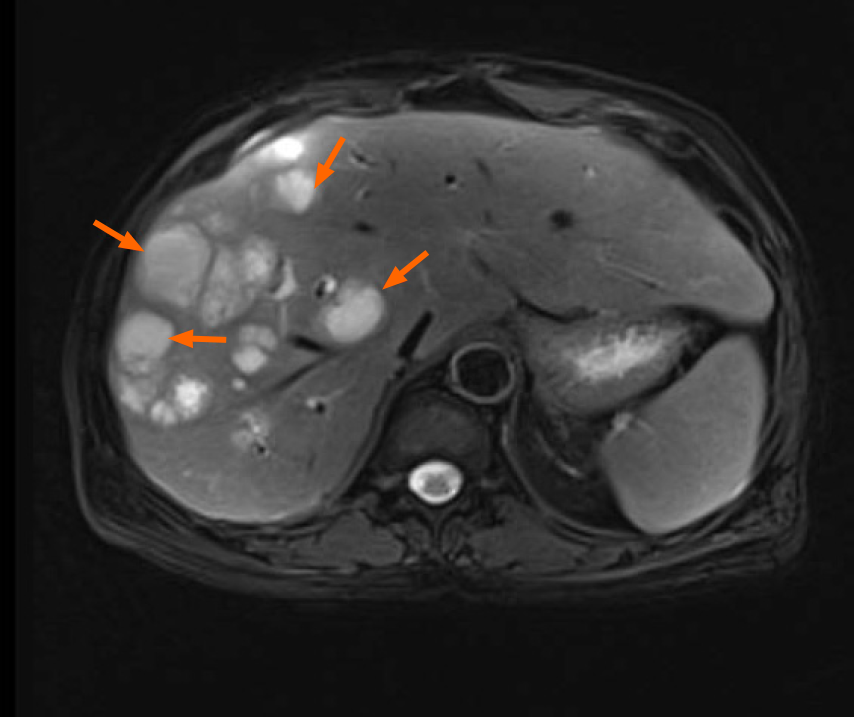
Figure 1 The liver lesions presented as long T1 and long T2 signals on magnetic resonance imaging.
There were multiple liver abscesses in right liver and perihepatic space.
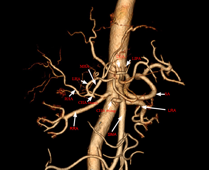
Figure 2 Computer tomography angiography postoperative.
Trifurcation of the celiac trunk was absent, and the left gastric artery and splenic artery originated in the anterior wall of the abdominal aorta, while the common hepatic artery (CHA) originated from the superior mesenteric artery. Unfortunately, the CHA was injured and the left subphrenic and middle hepatic arteries were dilated. In addition, the right hepatic artery supplied part of the right hepatic blood flow. Middle hepatic artery and left renal artery supplied the middle and left hepatic blood flow, respectively.
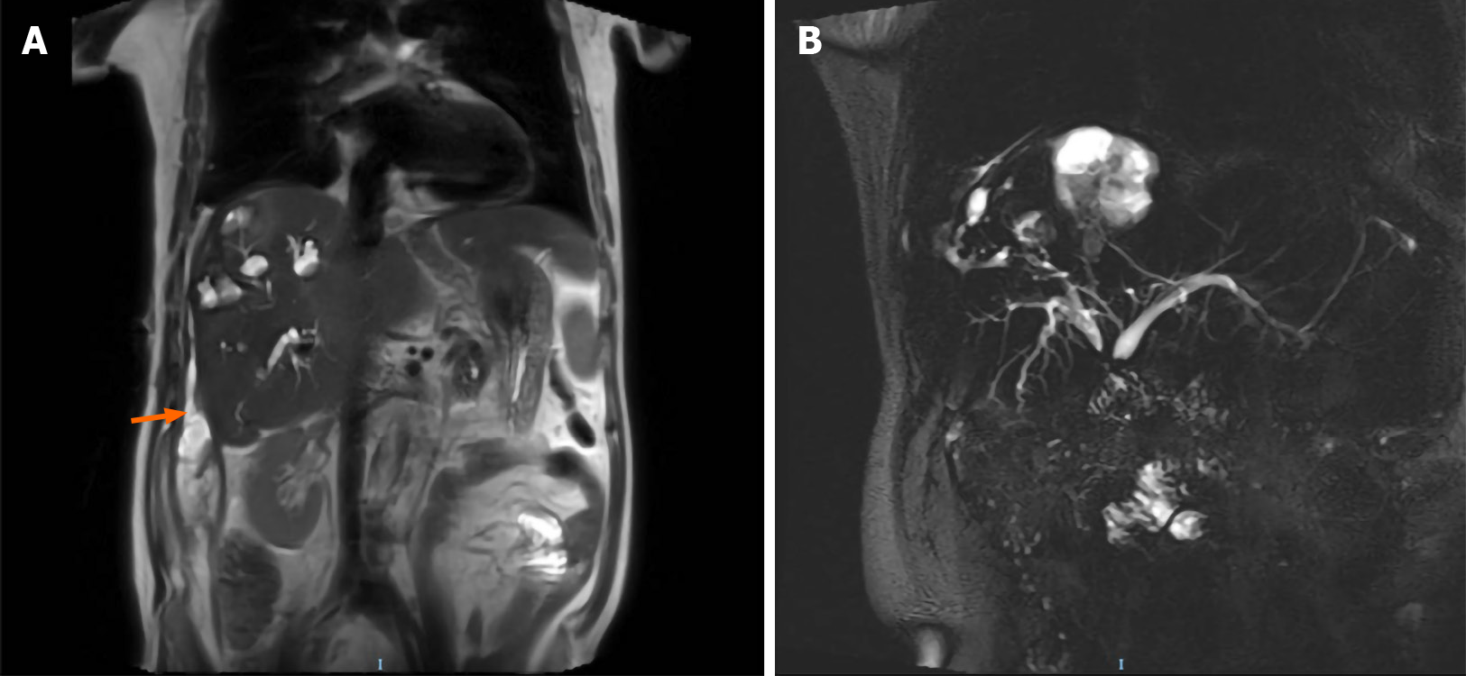
Figure 3 The image of magnetic resonance imaging in coronal section.
A: The results showed the liver abscesses were interlinked with the intrahepatic bile duct and the bile and pus flows to the perihepatic space; B: Magnetic resonance cholangiogram picture illustrated anatomical changes of bile duct after pancreatoduodenectomy.
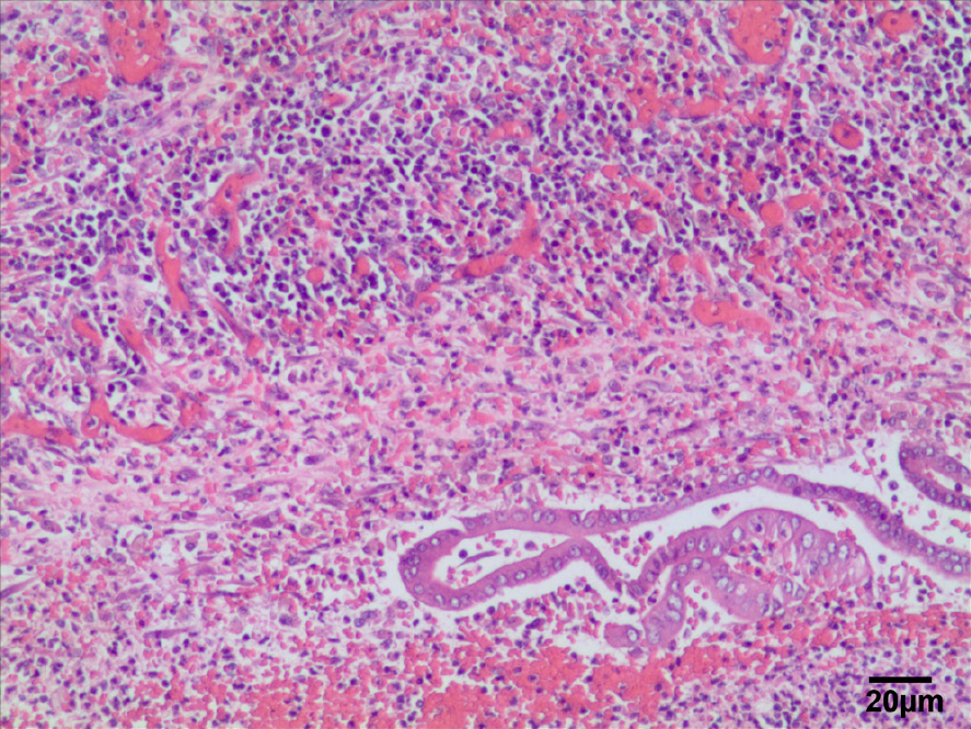
Figure 4 Histopathological examination of the liver.
Hematoxylin-eosin staining showed that there was necrotic liver tissue with liver degeneration and inflammatory cell infiltration. Scar bar: 20 μm.
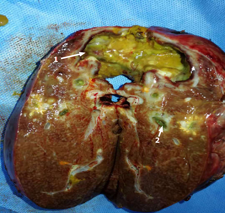
Figure 5 Resected specimen photo.
1The images displayed the liver abscess. 2Bile duct necrosis in the liver.
- Citation: Xie F, Wang J, Yang Q. Recurrent pyogenic liver abscess after pancreatoduodenectomy caused by common hepatic artery injury: A case report. World J Clin Cases 2021; 9(30): 9198-9204
- URL: https://www.wjgnet.com/2307-8960/full/v9/i30/9198.htm
- DOI: https://dx.doi.org/10.12998/wjcc.v9.i30.9198


