Copyright
©The Author(s) 2021.
World J Clin Cases. Jan 26, 2021; 9(3): 672-676
Published online Jan 26, 2021. doi: 10.12998/wjcc.v9.i3.672
Published online Jan 26, 2021. doi: 10.12998/wjcc.v9.i3.672
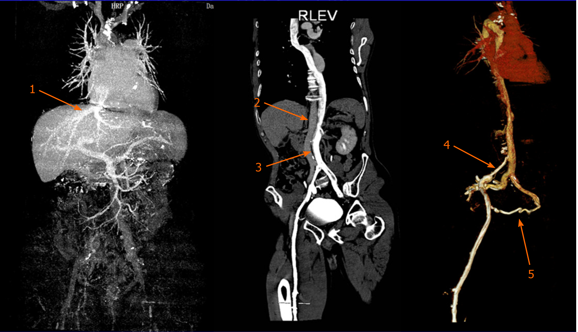
Figure 1 Contrast-enhanced computed tomography revealing a case of left-sided inferior vena cava draining into the hemiazygos vein with the hepatic vein directly draining into the atrium.
1: The hepatic vein directly draining into the atrium; 2: The abdominal aorta; 3: The left-sided inferior vena cava; 4: Right iliac vein stenosis; 5: Collateral vessels.
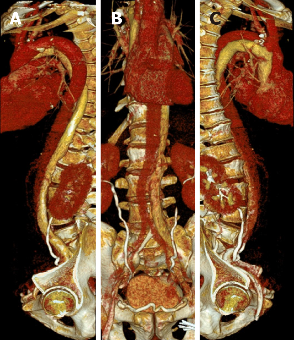
Figure 2 Three-dimensional reconstructed lateral and frontal views of the left inferior vena cava, vertebrae and the azygos venous system.
A: T8: 8th thoracic vertebra; B: T12: 12th thoracic vertebra; C: T5: 5th thoracic vertebra.
- Citation: Zhang L, Guan WK. Deep vein thrombosis in patient with left-sided inferior vena cava draining into the hemiazygos vein: A case report. World J Clin Cases 2021; 9(3): 672-676
- URL: https://www.wjgnet.com/2307-8960/full/v9/i3/672.htm
- DOI: https://dx.doi.org/10.12998/wjcc.v9.i3.672


