Copyright
©The Author(s) 2021.
World J Clin Cases. Sep 26, 2021; 9(27): 8274-8279
Published online Sep 26, 2021. doi: 10.12998/wjcc.v9.i27.8274
Published online Sep 26, 2021. doi: 10.12998/wjcc.v9.i27.8274
Figure 1 Changes in right-side facial expression muscle function before and after treatment.
A: Patient with a right facial droop before treatment; B and C: Patient with normal right-side facial expression muscle function after treatment.
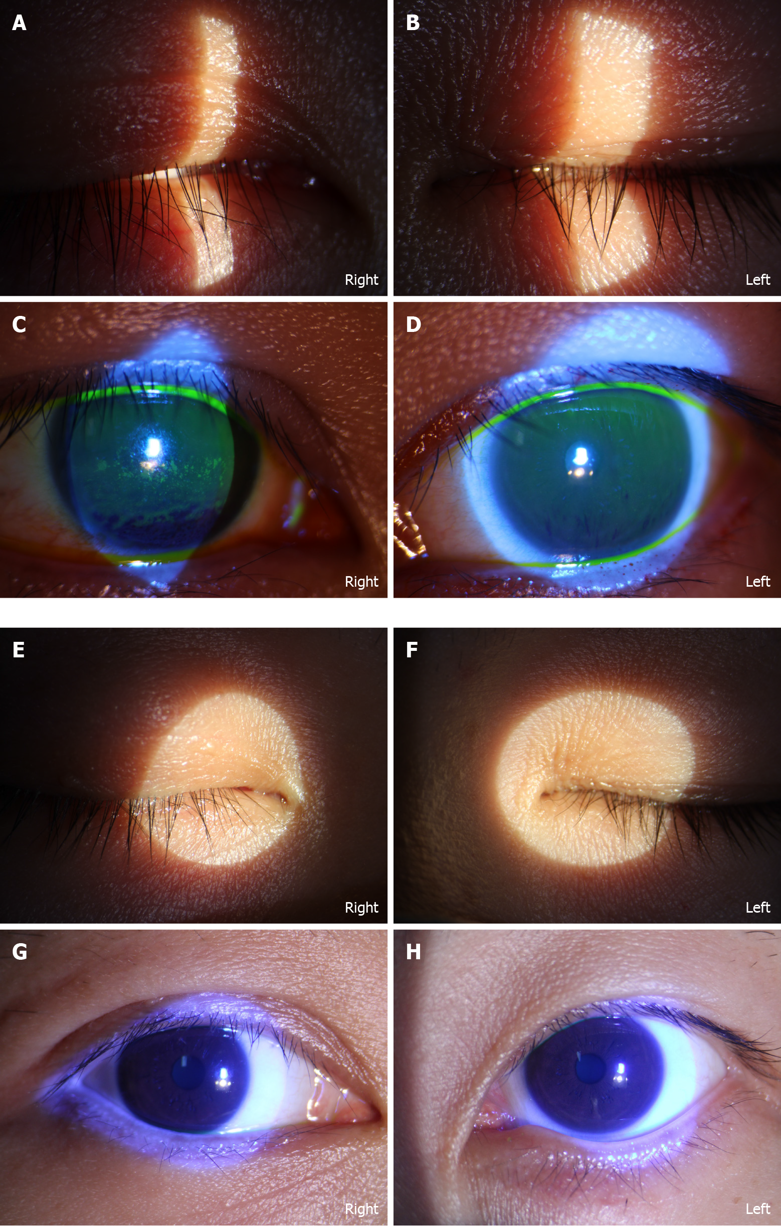
Figure 2 Ophthalmologic changes of both eyes before and after treatment.
A and B: Slit lamp inspection shows that the right eyelid could not completely close and that the left one could close normally; C and D: Conjunctival and scleral vessels are slightly congested. The central corneal epithelium of both eyes was punctate with opacity; E and F: Slit lamp inspection shows that both eyelids could close normally; G and H: Conjunctival and scleral vessels were not congested. Central corneal epithelium of both eyes had recovered.
- Citation: Yu BY, Cen LS, Chen T, Yang TH. Bell’s palsy after inactivated COVID-19 vaccination in a patient with history of recurrent Bell’s palsy: A case report. World J Clin Cases 2021; 9(27): 8274-8279
- URL: https://www.wjgnet.com/2307-8960/full/v9/i27/8274.htm
- DOI: https://dx.doi.org/10.12998/wjcc.v9.i27.8274


