Copyright
©The Author(s) 2021.
World J Clin Cases. Sep 26, 2021; 9(27): 8192-8198
Published online Sep 26, 2021. doi: 10.12998/wjcc.v9.i27.8192
Published online Sep 26, 2021. doi: 10.12998/wjcc.v9.i27.8192
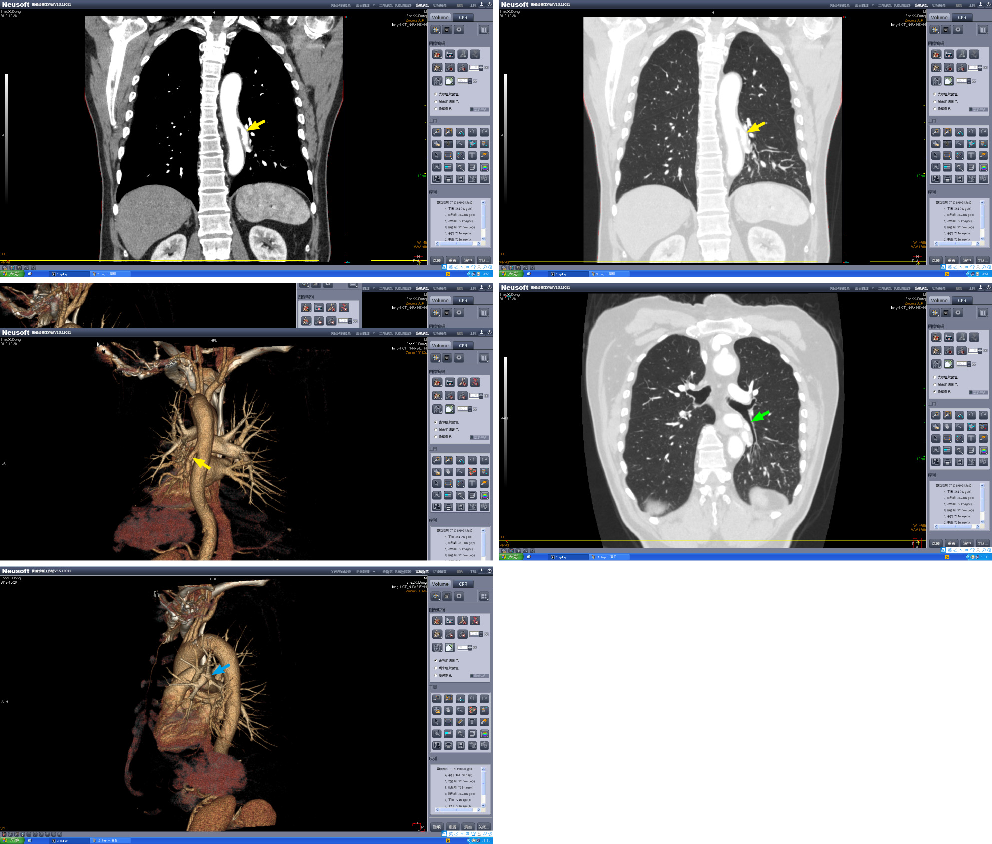
Figure 1 Contrast-enhanced computed tomography scan images with three-dimensional imaging.
The yellow arrows show the abnormal systemic artery. The green arrow shows the normal left inferior lobar bronchus. The blue arrow shows the left superior lobe pulmonary artery and its branches, and the left lower lobe pulmonary artery is missing.
- Citation: Zhang YY, Gu XY, Li JL, Liu Z, Lv GY. Surgical treatment of abnormal systemic artery to the left lower lobe: A case report . World J Clin Cases 2021; 9(27): 8192-8198
- URL: https://www.wjgnet.com/2307-8960/full/v9/i27/8192.htm
- DOI: https://dx.doi.org/10.12998/wjcc.v9.i27.8192


