Copyright
©The Author(s) 2021.
World J Clin Cases. Sep 26, 2021; 9(27): 8090-8096
Published online Sep 26, 2021. doi: 10.12998/wjcc.v9.i27.8090
Published online Sep 26, 2021. doi: 10.12998/wjcc.v9.i27.8090
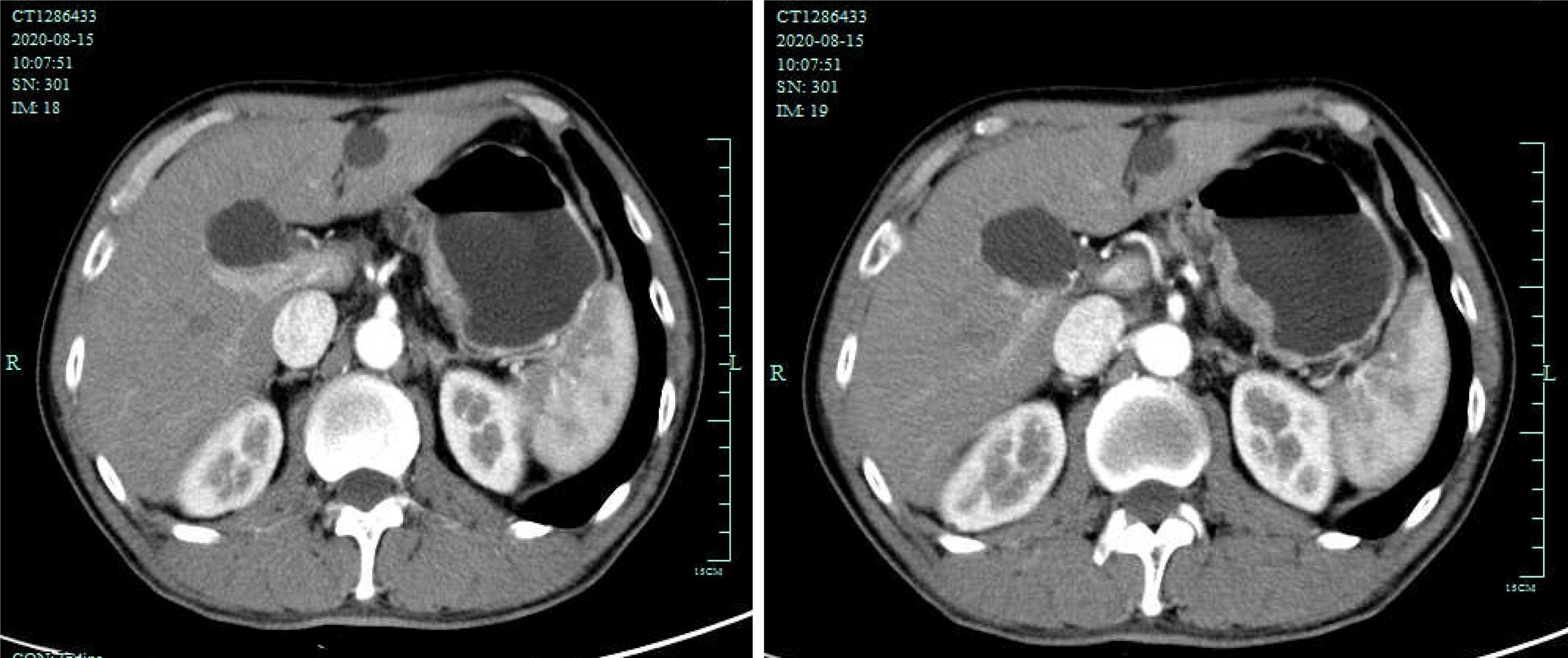
Figure 1 Preoperation enhanced abdominal computed tomography images.
The images revealed the gastric wall at the lesser curvature of remnant stomach were obviously thickened. No enlarged lymph nodes were evident around the stomach.
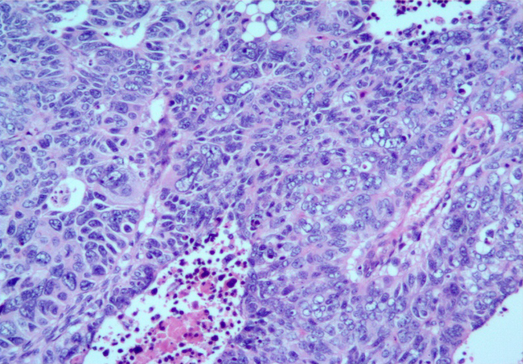
Figure 2 Histological findings.
Poorly differentiated carcinoma tumor tissues with necrosis (hematoxylin and eosin × 200).
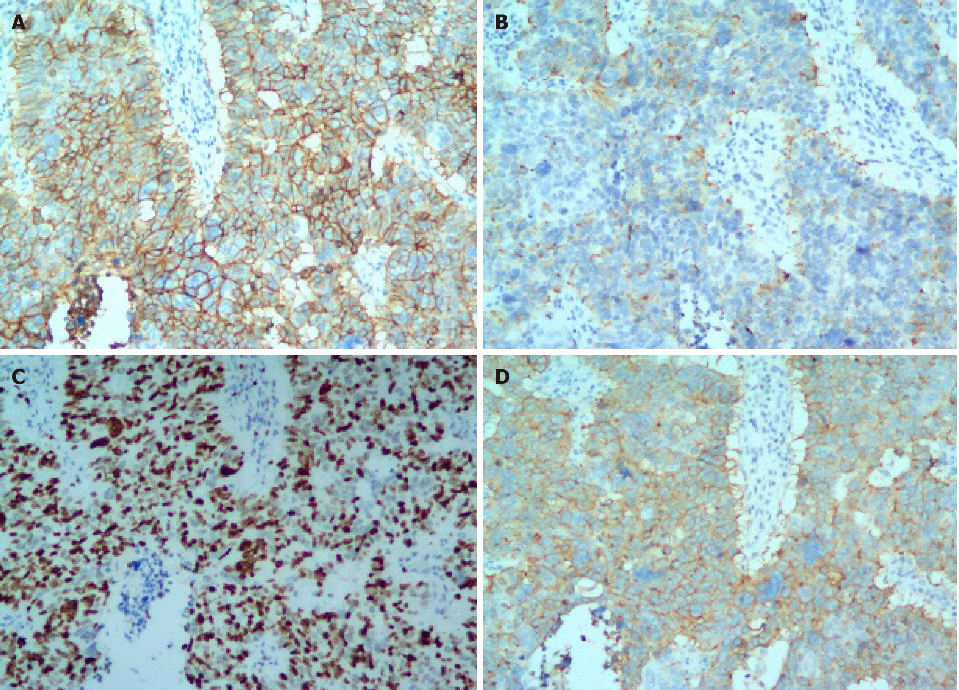
Figure 3 Immunohistochemical staining.
A: Positive immunohistochemical staining for CD56 (× 200); Positive immunohistochemical staining for chromogranin A (× 200); The Ki-67 index was about 80% (× 200); Positive staining for synaptophysin (× 200).
- Citation: Zhu H, Zhang MY, Sun WL, Chen G. Mixed neuroendocrine carcinoma of the gastric stump: A case report. World J Clin Cases 2021; 9(27): 8090-8096
- URL: https://www.wjgnet.com/2307-8960/full/v9/i27/8090.htm
- DOI: https://dx.doi.org/10.12998/wjcc.v9.i27.8090


