Copyright
©The Author(s) 2021.
World J Clin Cases. Jun 6, 2021; 9(16): 4090-4094
Published online Jun 6, 2021. doi: 10.12998/wjcc.v9.i16.4090
Published online Jun 6, 2021. doi: 10.12998/wjcc.v9.i16.4090
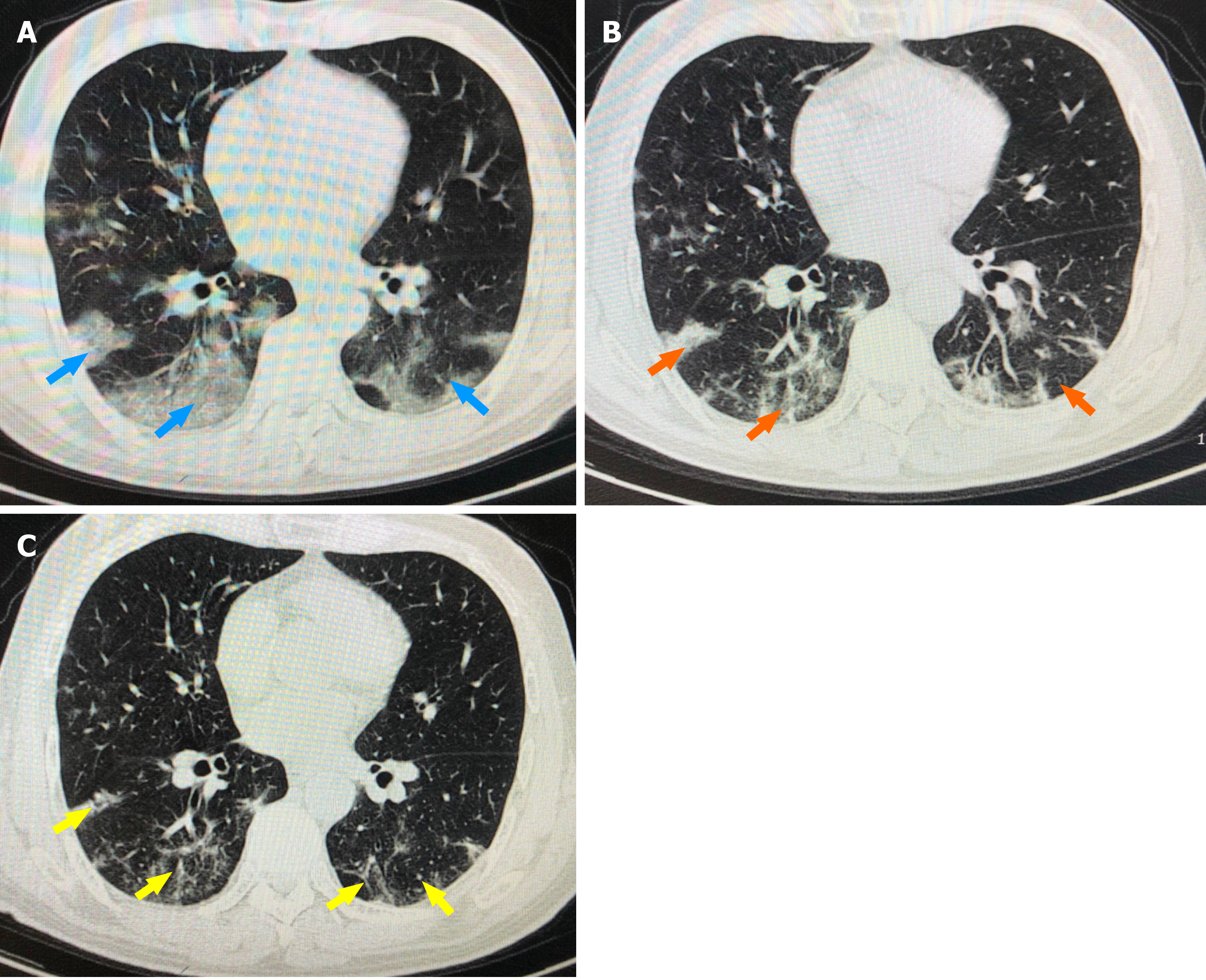
Figure 1 Chest computed tomography results of the coronavirus disease 2019 patient.
A: Computed tomography (CT) of the chest on April 28. Blue arrows show multiple patchy shadows and ground glass shadows in both lungs; B: CT of the chest on May 1. Orange arrows show obvious absorption of the lesions in both lungs; C: CT of the chest on May 11. Yellow arrows show that the lesions in both lungs were absorbed.
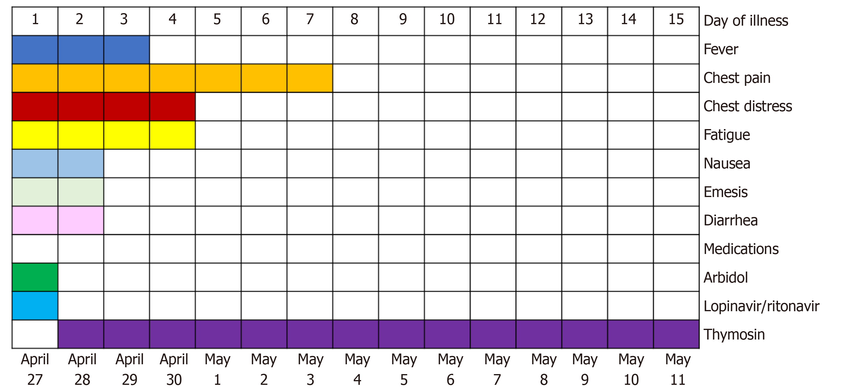
Figure 2 Symptoms and treatments of the patient outlined accordingly to days post symptom onset since April 27, 2020.
- Citation: Zheng QN, Xu MY, Gan FM, Ye SS, Zhao H. Thymosin as a possible therapeutic drug for COVID-19: A case report. World J Clin Cases 2021; 9(16): 4090-4094
- URL: https://www.wjgnet.com/2307-8960/full/v9/i16/4090.htm
- DOI: https://dx.doi.org/10.12998/wjcc.v9.i16.4090


