Copyright
©The Author(s) 2021.
World J Clin Cases. Jun 6, 2021; 9(16): 4062-4071
Published online Jun 6, 2021. doi: 10.12998/wjcc.v9.i16.4062
Published online Jun 6, 2021. doi: 10.12998/wjcc.v9.i16.4062
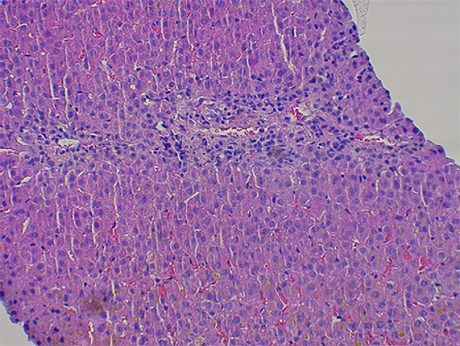
Figure 1 The liver biopsy specimen of case 1.
Hematoxylin and eosin-stained histological evidence showing cholestatic injury, ductopenia, and chronic hepatitis with mild fibrosis after ligandrol and post-cycle therapy misuse. In the lobules, there is predominantly canalicular cholestasis, dilation and numerous biliary plugs between hepatocytes, in the Kupffer cells and fewer in the cytoplasm of centrilobular hepatocytes. There are focal degenerative signs of hepatocytes with vacuolization, without steatosis, with some spotty necrosis. In the portobiliary areas, there is a moderately dense inflammatory infiltrate with lymphocytes cluster of differentiation 3+ (CD3+), CD20 focally+, CD138 +/-, sketchy mild interface hepatitis, ductopenia, almost complete loss of biliary ducts, and peripheral ductular metaplasia of periportal hepatocytes (cytokeratin7+). Mild mononuclear inflammatory infiltrates in the sinusoids, markedly multiplied Kupffer cells with cholestasis. Mild portal fibrosis (Dr. Meciarova).
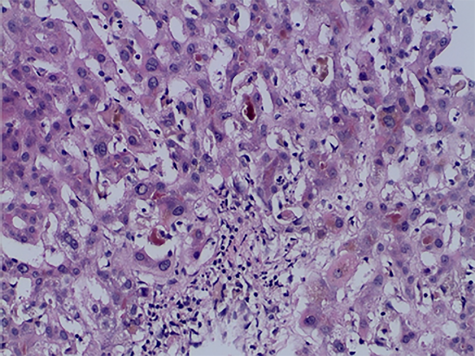
Figure 2 The liver biopsy specimen of case 2.
Hematoxylin and eosin-stained histological evidence showing acute cholestasis with mild fibrosis after ligandrol and post-cycle therapy misuse. Hepatocyte architecture is preserved without nodularity with some hepatocyte apoptosis and perivenular hepatocyte necrosis, or patches of swollen hepatocytes with double nuclei. No apparent signs of significant steatosis, Mallory-Denk bodies, or hemosiderin content. Porto-biliary areas have distinct band-like enlargement, with some thin threads of bridging fibrosis together with destruction of bile ducts with anisokaryosis and focally overlapping lines of biliary epithelial cell nuclei with ductular reaction and a mixed inflammatory infiltrate with focal neutrophil content. There is a marked centroacinar canalicular cholestasis with numerous bile-plugs in the canaliculi with abundant phagocytosis by the Kupffer cells (Schmorl reaction). Immunohistochemistry stains: cytokeratin 7+ in the bile ducts, ductules, and robust biliary metaplasia, cytokeratin 8/18+ and factor VIII in endothelial cells, cluster of differentiation 34+ (CD34+) in the periportal capilarized sinusoids, ubiquitin glutamine synthetase + in the centroacinar foci, CD15+ in neutrophils and macrophages, and LCA in the lymphocytes (Dr. Smitka).
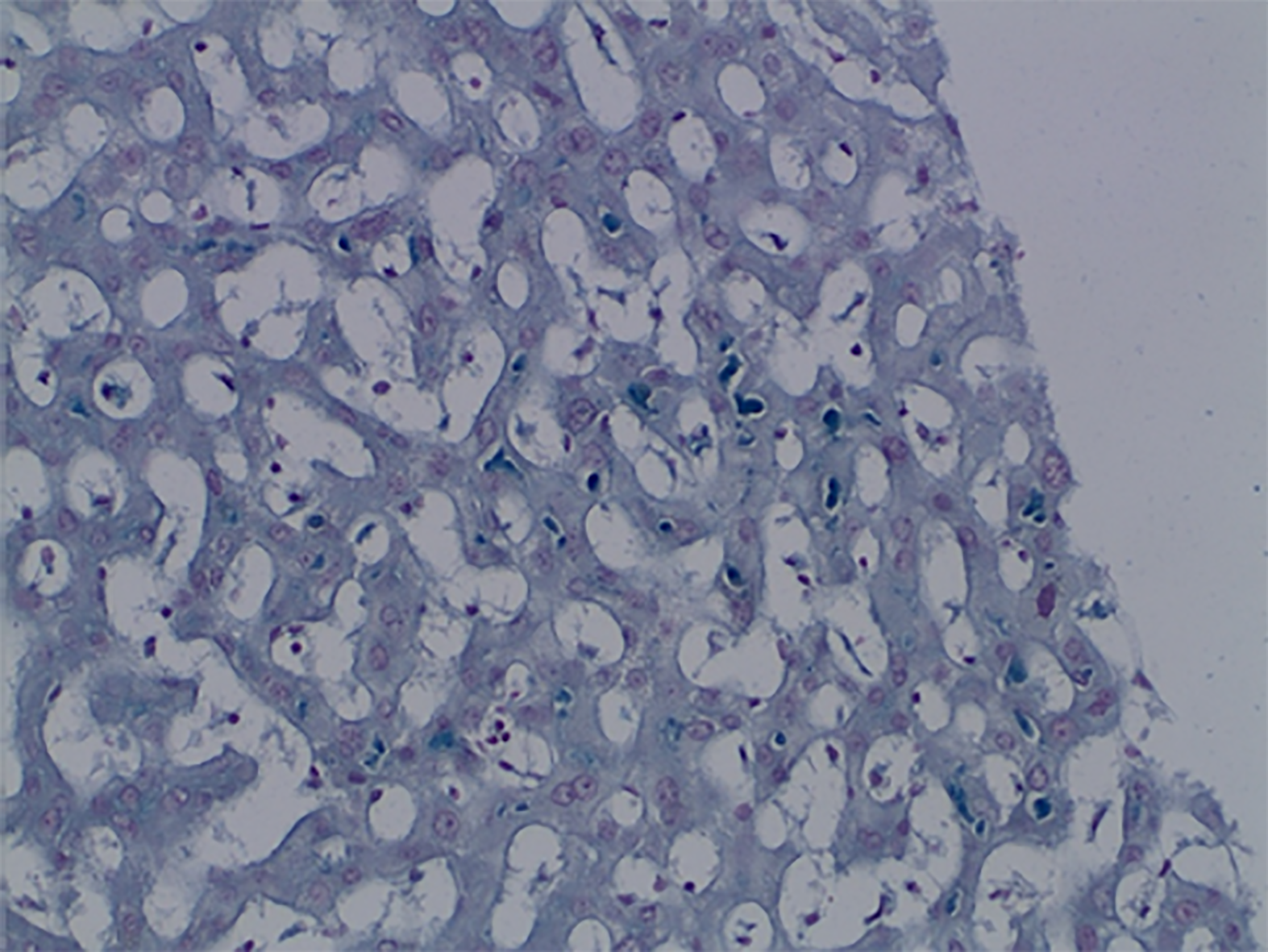
Figure 3 The liver biopsy specimen of case 2 patient, special staining.
Schmorl stained histological evidence showing acute cholestasis after ligandrol and post-cycle therapy misuse. Deposition of blue-stained bile salts in various locations in the liver parenchyma (Dr. Smitka).
- Citation: Koller T, Vrbova P, Meciarova I, Molcan P, Smitka M, Adamcova Selcanova S, Skladany L. Liver injury associated with the use of selective androgen receptor modulators and post-cycle therapy: Two case reports and literature review . World J Clin Cases 2021; 9(16): 4062-4071
- URL: https://www.wjgnet.com/2307-8960/full/v9/i16/4062.htm
- DOI: https://dx.doi.org/10.12998/wjcc.v9.i16.4062


