Copyright
©The Author(s) 2020.
World J Clin Cases. Apr 6, 2020; 8(7): 1301-1305
Published online Apr 6, 2020. doi: 10.12998/wjcc.v8.i7.1301
Published online Apr 6, 2020. doi: 10.12998/wjcc.v8.i7.1301
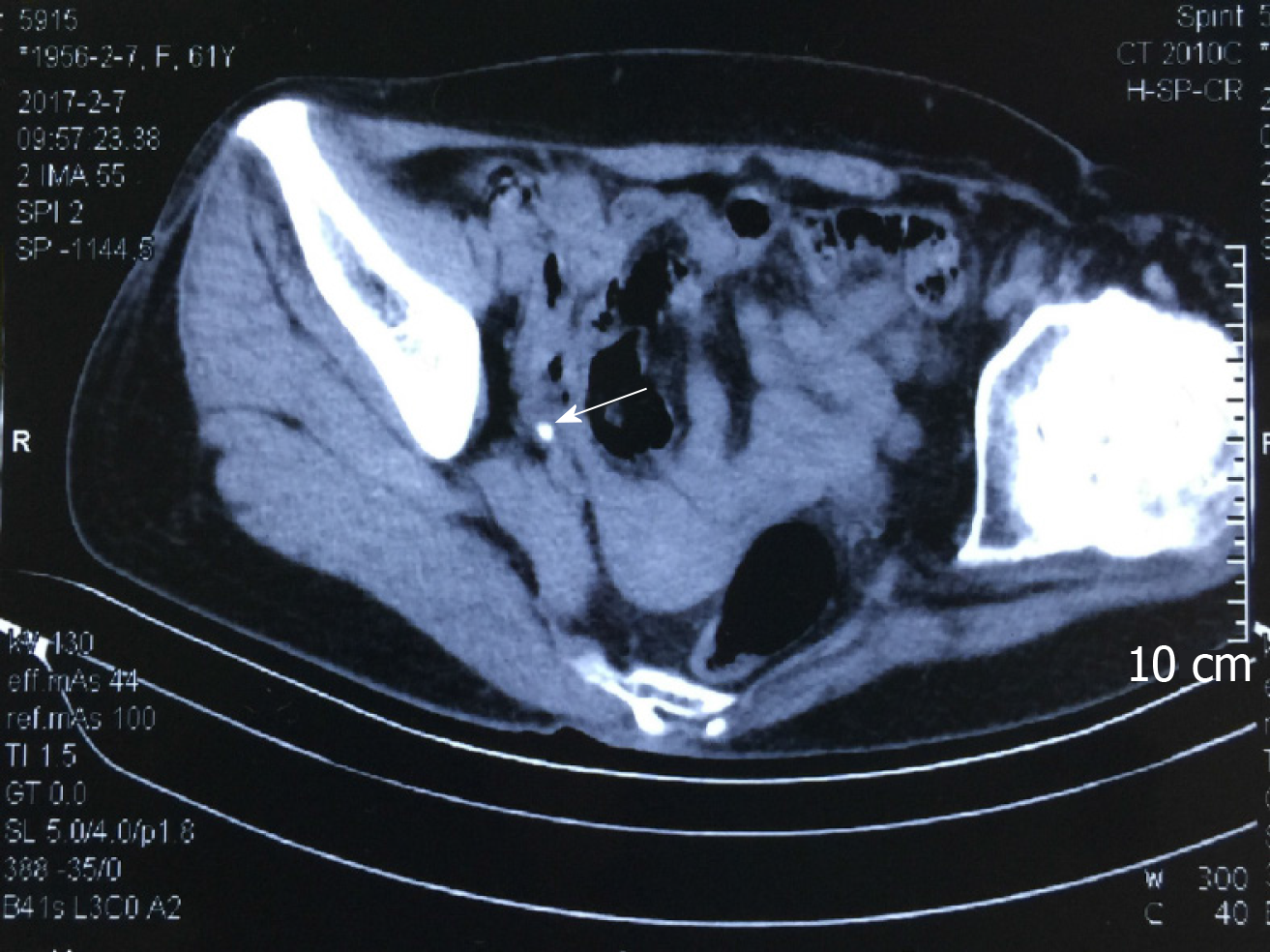
Figure 1 Computed tomography scan showing a calculi in the right ureter.
Arrowhead indicates the location of ureteral calculi.
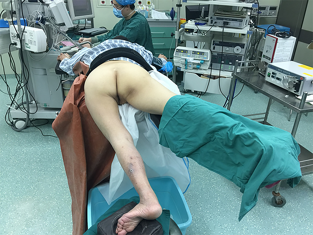
Figure 2 Patient positioning in the modified prone split-leg position due to limited movement of her left hip.
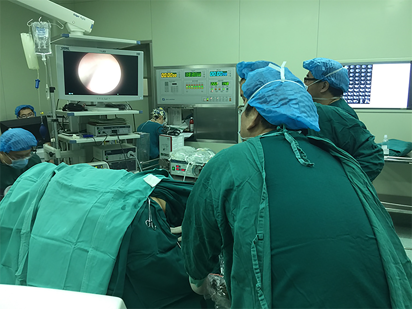
Figure 3 Ureteroscope enters into the right ureter alongside the guide wire.
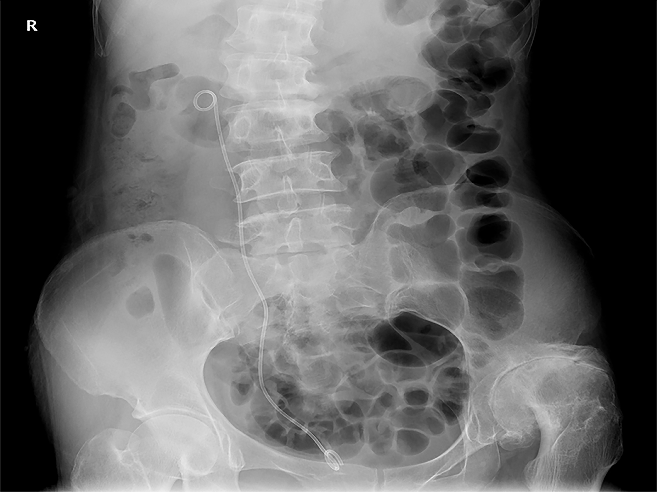
Figure 4 X-ray of kidney, ureter, and bladder shows stent position and no visible stone in ureteral area after operation.
- Citation: Huang K. Rigid ureteroscopy in prone split-leg position for fragmentation of female ureteral stones: A case report. World J Clin Cases 2020; 8(7): 1301-1305
- URL: https://www.wjgnet.com/2307-8960/full/v8/i7/1301.htm
- DOI: https://dx.doi.org/10.12998/wjcc.v8.i7.1301


