Copyright
©The Author(s) 2020.
World J Clin Cases. Apr 6, 2020; 8(7): 1203-1212
Published online Apr 6, 2020. doi: 10.12998/wjcc.v8.i7.1203
Published online Apr 6, 2020. doi: 10.12998/wjcc.v8.i7.1203
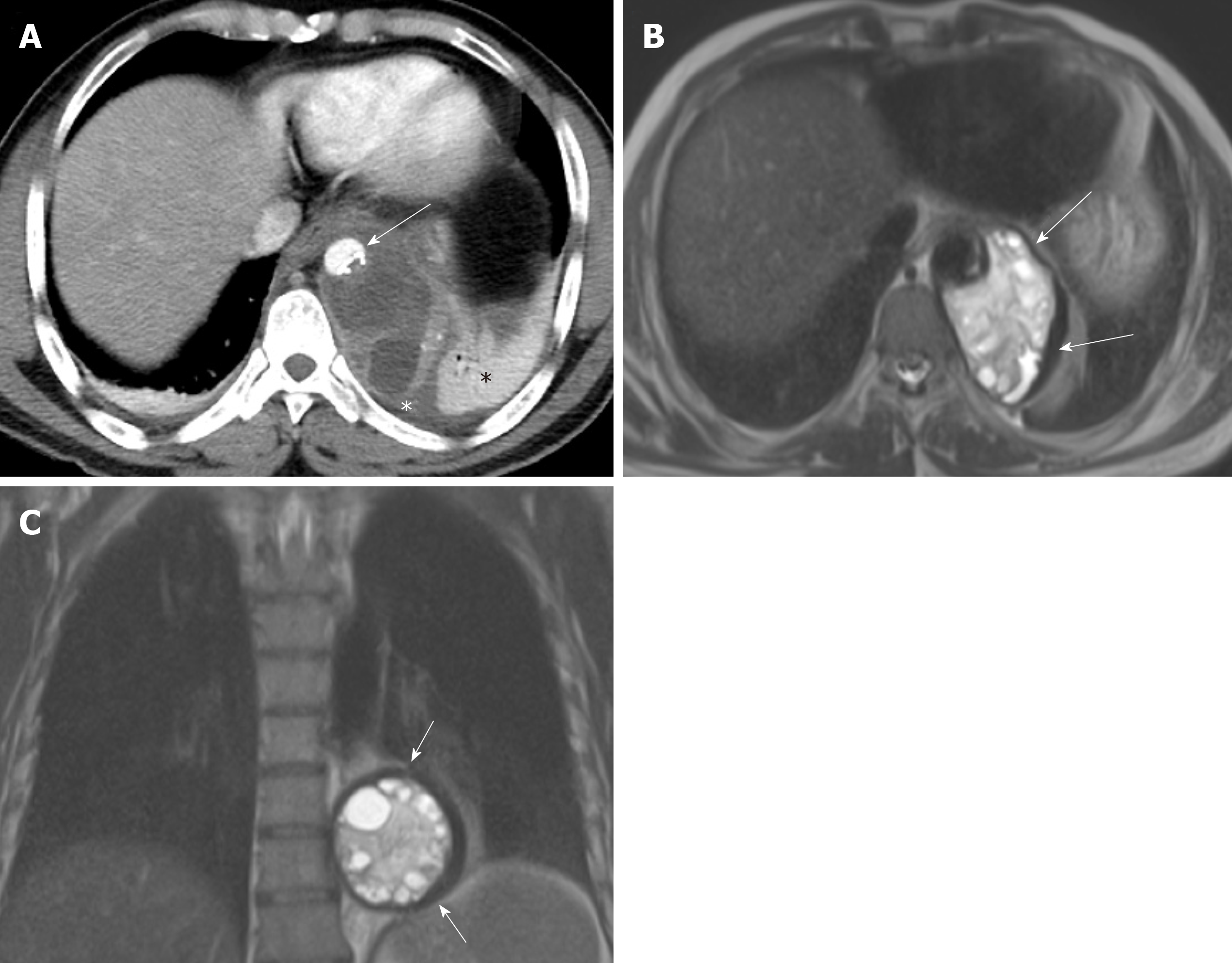
Figure 1 A 48-year old male with mediastinal hydatid cyst presented with acute chest pain.
A: Axial image of enhanced computed tomography angiogram shows a complex cystic lesion with thick wall (white arrows) that is predominately located in the left side of the posterior mediastinum with evidence small calcifications at the periphery of the lesions at the site of abutment with descending thoracic aorta. There is adjacent pleural effusion (white asterisk) and enhancing left lower lobe atelectasis (white asterisk); B, C: Axial and coronal T2 weighted images show a complex cystic lesion with thick dark rim (arrows) abutting the descending thoracic aorta with multiple peripheral daughter cysts of higher signal intensity compared to the intermediately hyperintenese matrix of the mother cyst. Magnetic resonance imaging findings are highly suggestive of hydatid cyst, likely primary given absence of evidence of lung and liver hydatid disease.
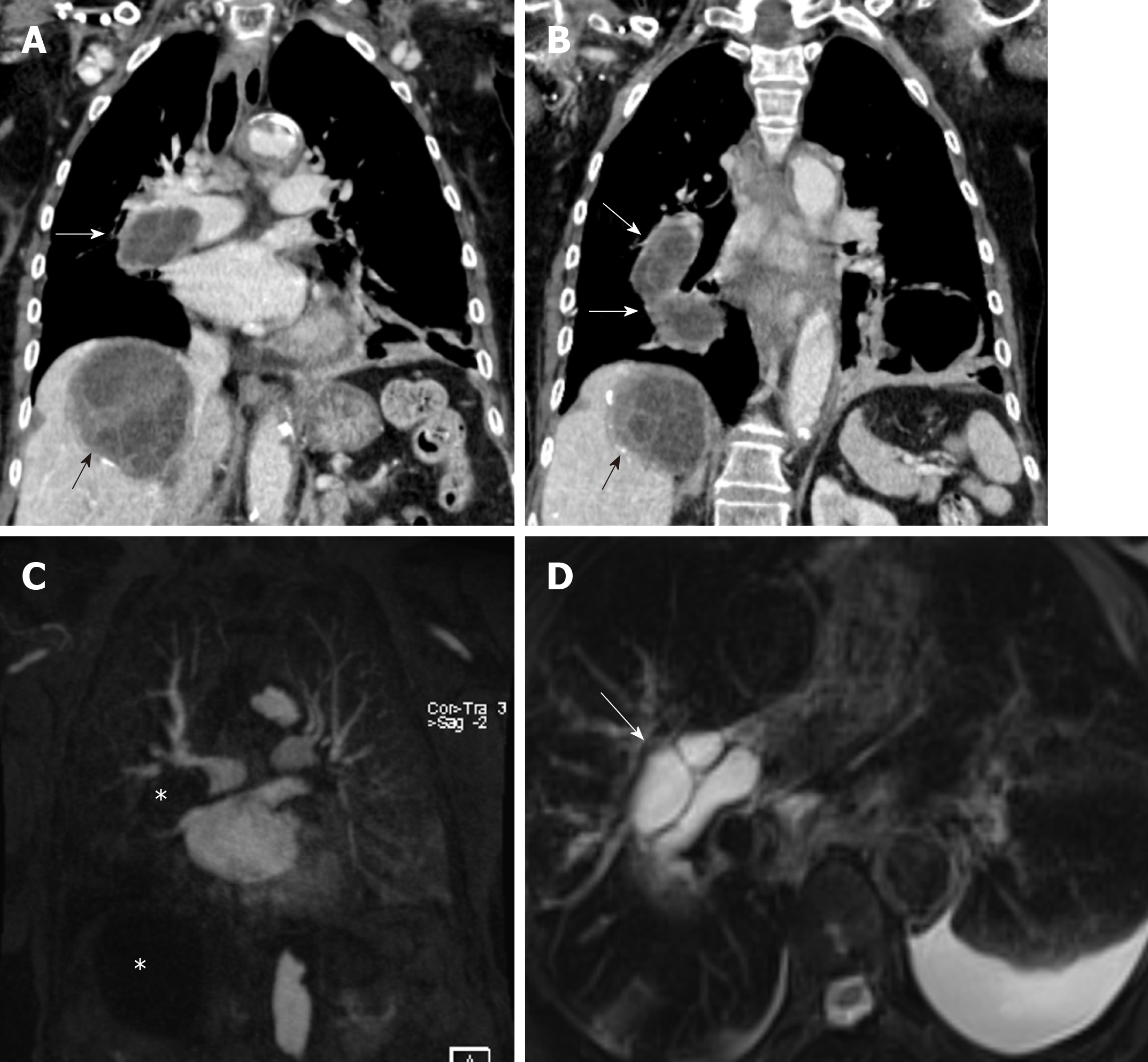
Figure 2 An 82-year-old female with pulmonary artery hydatid cyst embolism presented with shortness of breath.
A, B: Coronal images of enhanced computed tomography scan show right main pulmonary artery intra-luminal cystic filling defects causing expansion and near occlusion of the artery with extension into the right interlobar pulmonary artery (white arrows). There is hepatic hydatid cyst with internal septation and daughter cysts and scattered wall calcifications (black arrows); C: Coronal image of T1 weighted magnetic resonance angiography confirms the presence of filling defect of low signal intensity within the right pulmonary artery and low signal intensity liver lesion (asterisks) with no evidence of contrast enhancement; D: Axial T2 weighted image with fat saturation shows right main pulmonary artery intra-luminal cystic filling defects of high signal intensity with septations suggestive of daughter cysts (arrow). Left pleural effusion is noted.
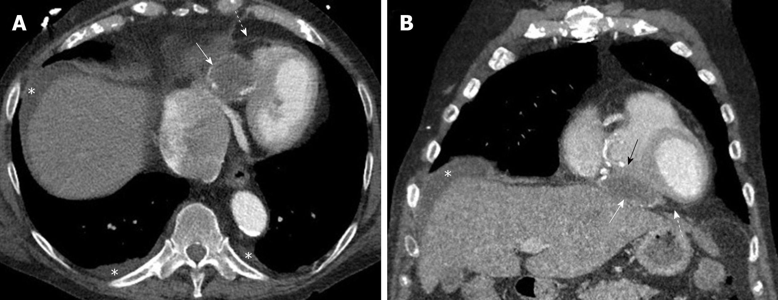
Figure 3 Pericardial hydatid cyst.
A, B: Axial and coronal contrast enhanced computed tomography images show a partially peripherally calcified cystic lesion (white arrows) along the inferior aspect of the heart and within the pericardial sac (dashed arrows) with mild mass effect and displacement of the right ventricle (black arrow). Small pleural effusions and ascites are shown (asterisks).
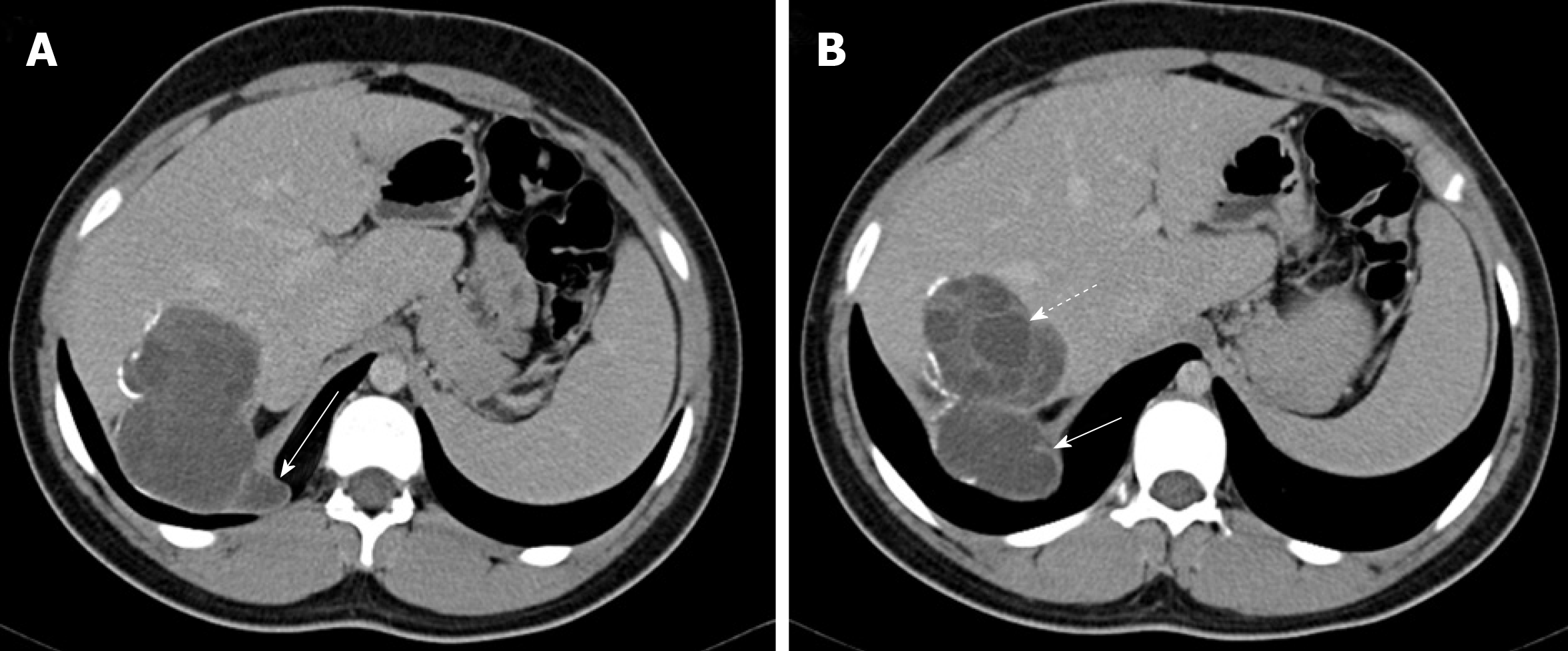
Figure 4 Hepatic hydatid cyst complicated by trans-diaphragmatic rupture in a 17 year old male patient.
A, B: Axial contrast enhanced computed tomography images show a segment 7 cyst with scattered wall calcifications and multiple daughter cysts (dashed arrow). There is cyst perforation through the right hemi-diaphragm with little invasion of the thoracic cavity (arrows).
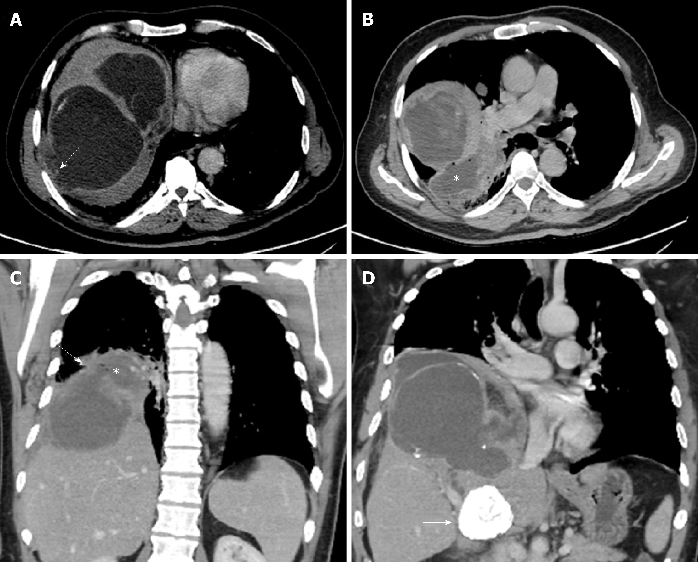
Figure 5 A 42-year old male presented with liver hydatid cyst complicated by broncho-biliary fistula presented with coughing bile.
Axial and coronal computed tomography scan images using liver window (A) and standard soft tissue window (B-D) show intercommunicating multiple liver cystic lesions with internal non-enhancing septations and peripheral calcifications within hepatic segments 8 and 7. The right lower lobe lung cystic lesion (asterisks) contains multiple air foci indicating bronchial communication with evidence of wide communication with the hepatic cystic lesion through the diaphragmatic defect (dashed arrows in A and C). A calcified hepatic lesion near the porta hepatis represents a healed hydatid cyst (arrow in D).
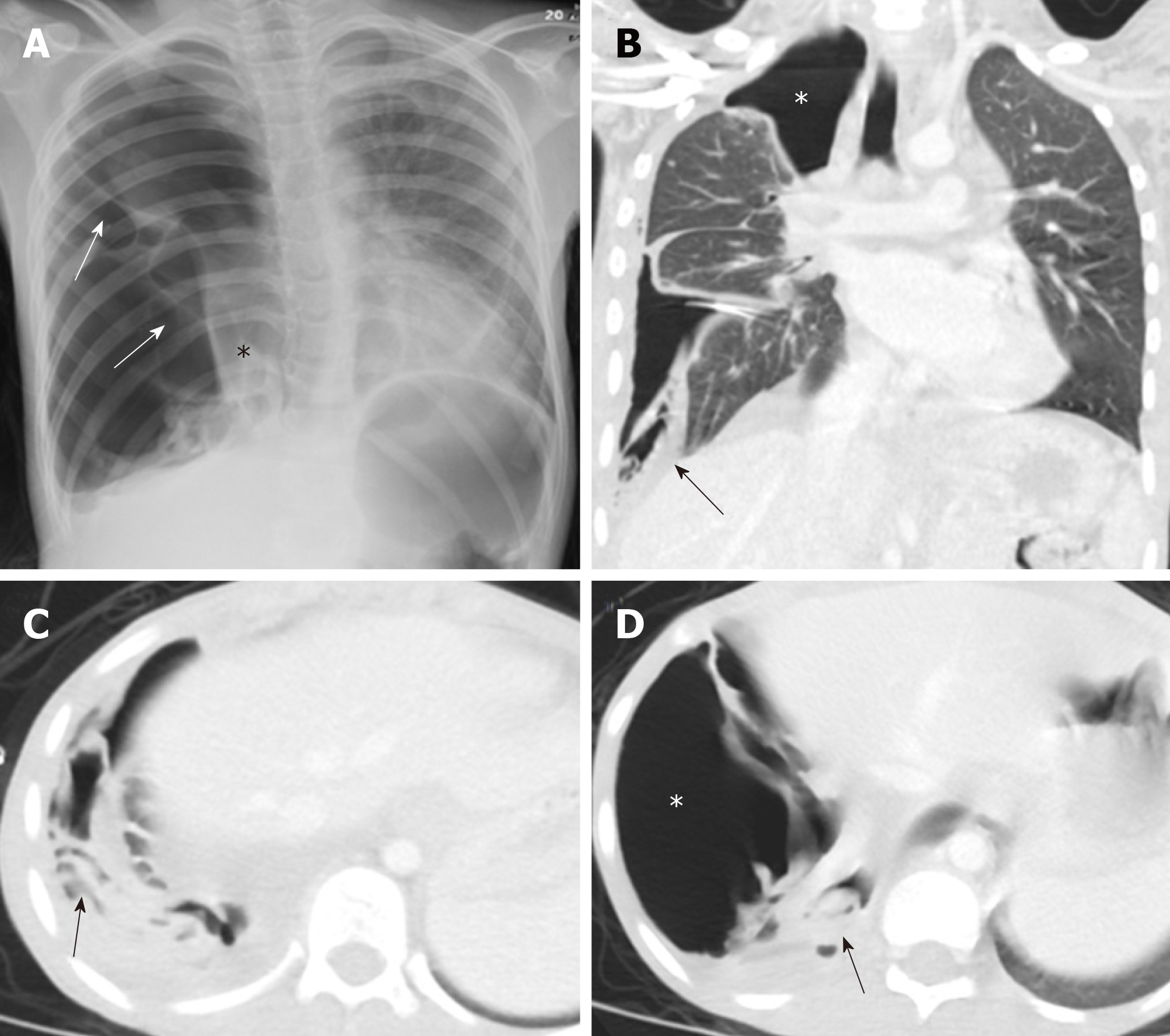
Figure 6 A 10-year old female with spontaneous ruptured lung hydatid cyst presenting with pneumothorax and complicated by broncho-pleural fistula.
Patient presented with oxygen desaturation and chest pain with chronic history of shortness of breath and cough for 3 months. A: Frontal chest radiograph shows large right-sided pneumothorax with small pleural effusion and multiple pleural septations (arrows), medially displaced collapsed right lung (asterisk), mild contralateral shift of the mediastinum to the left, and flattening of the right hemidiaphragm; B-D: Coronal and axial images of chest computed tomography scan after placement of chest tube show improvement of lung aeration with persistent large right-sided pneumothorax (asterisks) which suggest broncho-plural fistula, multiple collapsed membranes (arrows), and adjacent plural effusion.
- Citation: Saeedan MB, Aljohani IM, Alghofaily KA, Loutfi S, Ghosh S. Thoracic hydatid disease: A radiologic review of unusual cases. World J Clin Cases 2020; 8(7): 1203-1212
- URL: https://www.wjgnet.com/2307-8960/full/v8/i7/1203.htm
- DOI: https://dx.doi.org/10.12998/wjcc.v8.i7.1203


