Copyright
©The Author(s) 2020.
World J Clin Cases. Mar 26, 2020; 8(6): 1116-1128
Published online Mar 26, 2020. doi: 10.12998/wjcc.v8.i6.1116
Published online Mar 26, 2020. doi: 10.12998/wjcc.v8.i6.1116
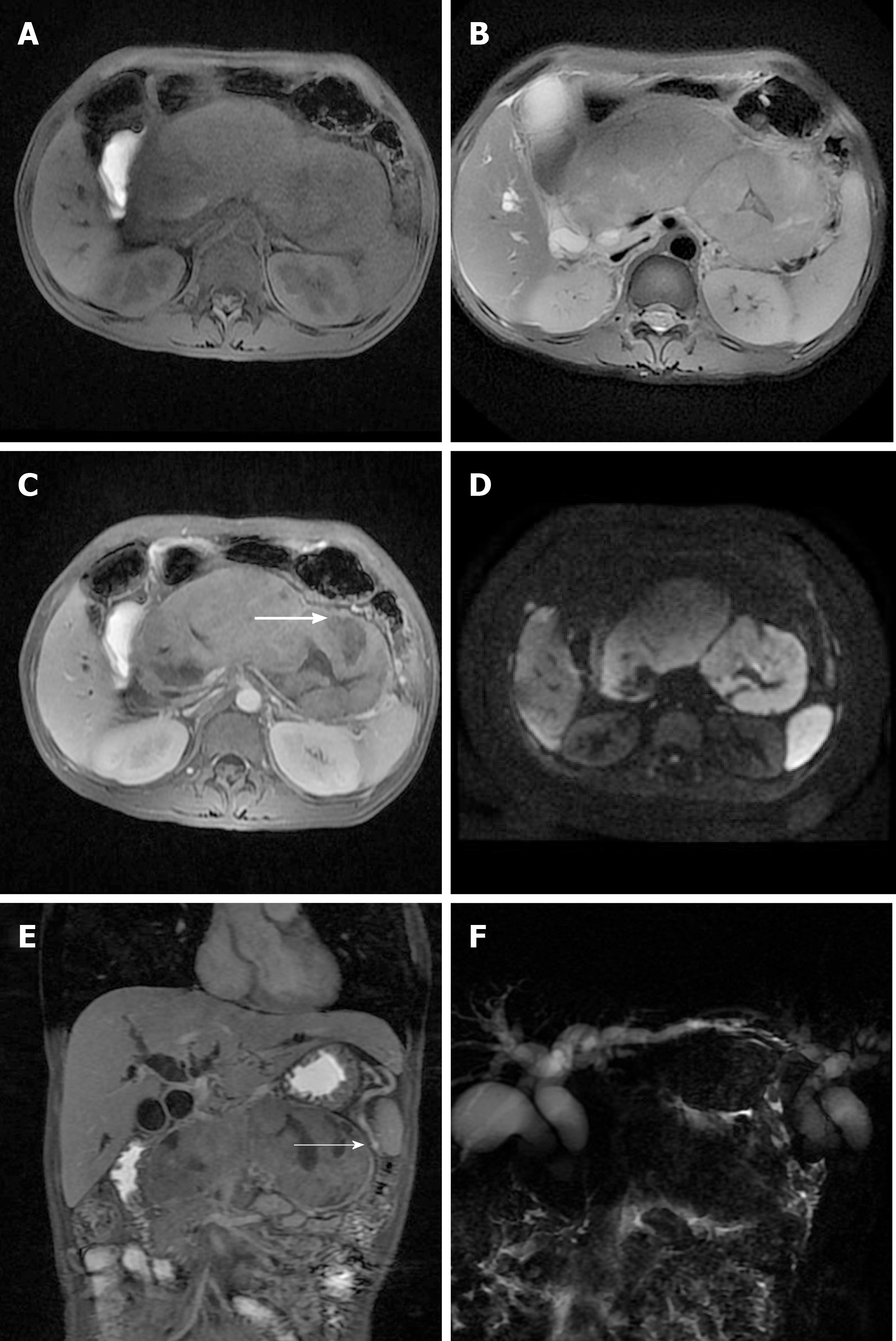
Figure 1 Magnetic resonance imaging of the patient.
Magnetic resonance imaging of the upper abdomen shows a diffusely enlarged pancreas, appearing “sausage-shaped”, with loss of pancreatic lobular structure. A: The lesion was isointense to hypointense in the fat-suppressed T1-weighted image; B: The lesion was isointense to hyperintense in the fat-suppressed T2-weighted image; C and D: Central necrosis (see thick arrow) was seen during the contrast-enhanced delayed phase and mixed signal intensity on DWI; E: In addition, there is a distinct hyperintense rim (see the thin arrow) that demonstrated delayed enhancement and a capsule appearance; F: Magnetic resonance cholangiopancreatography showed upstream dilation of the proximal common bile duct and intrahepatic biliary ducts secondary to strictures of the main pancreatic duct and distal common duct.
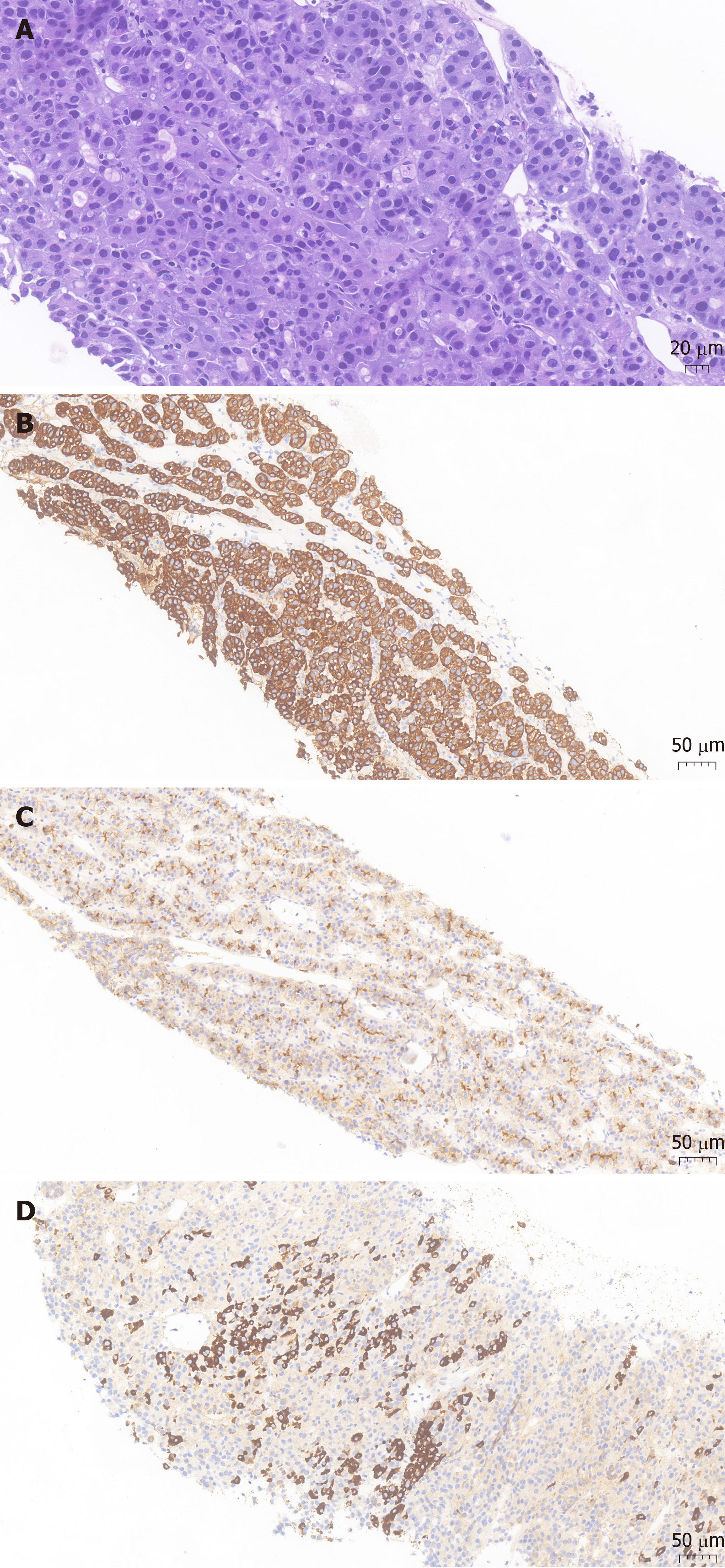
Figure 2 Pathological presentation of the patient.
A: Hematoxylin-eosin staining revealed heteromorphic neoplastic cells arranged in glandular, nested or striped patterns (magnification: × 200; scale bar: 20 μm); B: Immunohistochemistry showed tumor cell positivity for CK19 (magnification: × 100; scale bar: 50 μm); C: Immunohistochemistry showed tumor cell positivity for polyclonal carcinoembryonic antigen (magnification: × 100; scale bar: 50 μm); D: Immunohistochemistry showed tumor cell positivity for HepPar1 (magnification: × 200; scale bar: 50 μm).
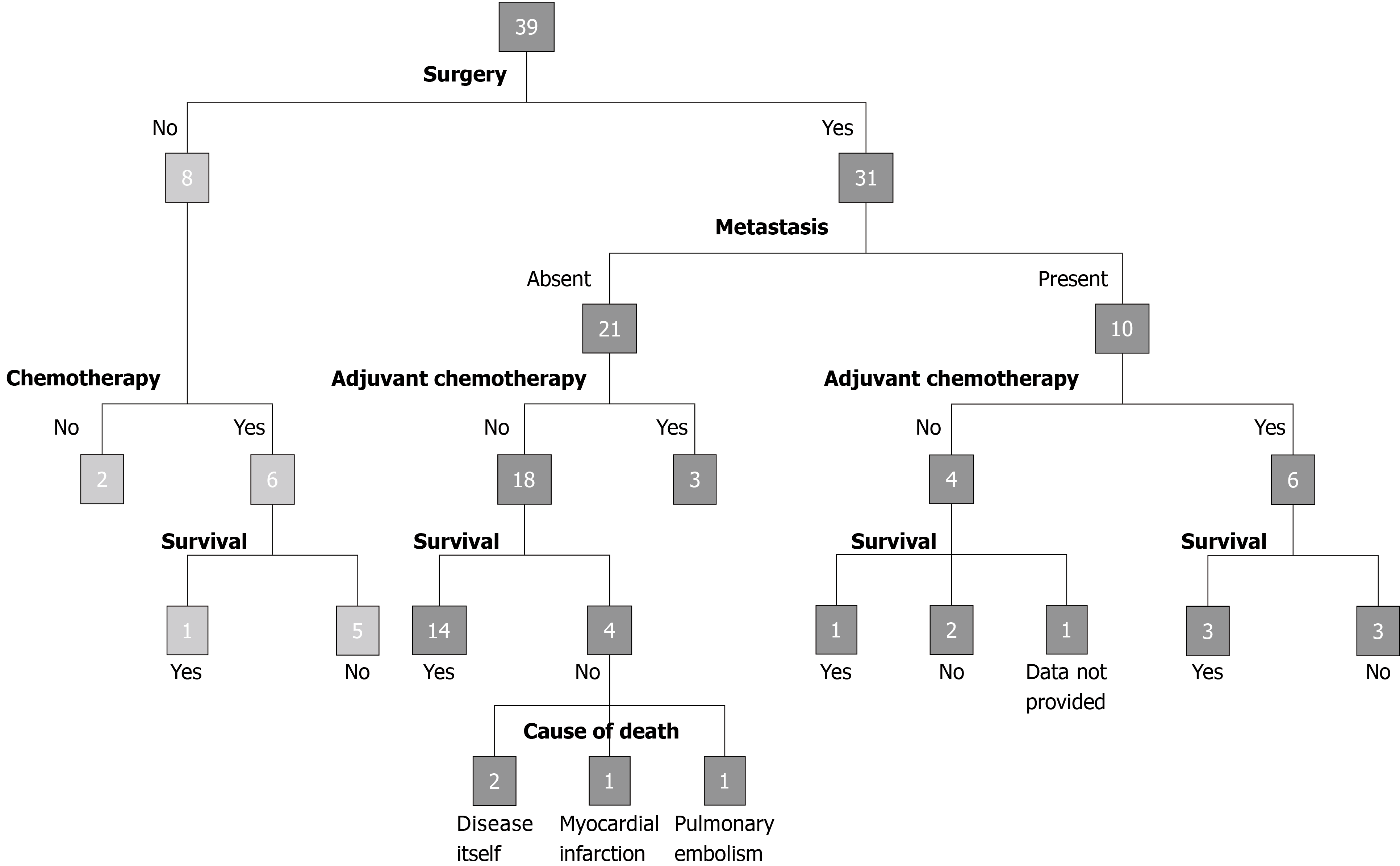
Figure 3 Outcomes of the 39 patients with hepatoid carcinoma of the pancreas reported in the literature.
- Citation: Zeng SX, Tan SW, Fong CJTH, Liang Q, Zhao BL, Liu K, Guo JX, Tao J. Hepatoid carcinoma of the pancreas: A case report and review of the literature. World J Clin Cases 2020; 8(6): 1116-1128
- URL: https://www.wjgnet.com/2307-8960/full/v8/i6/1116.htm
- DOI: https://dx.doi.org/10.12998/wjcc.v8.i6.1116


