Copyright
©The Author(s) 2020.
World J Clin Cases. Feb 26, 2020; 8(4): 798-805
Published online Feb 26, 2020. doi: 10.12998/wjcc.v8.i4.798
Published online Feb 26, 2020. doi: 10.12998/wjcc.v8.i4.798
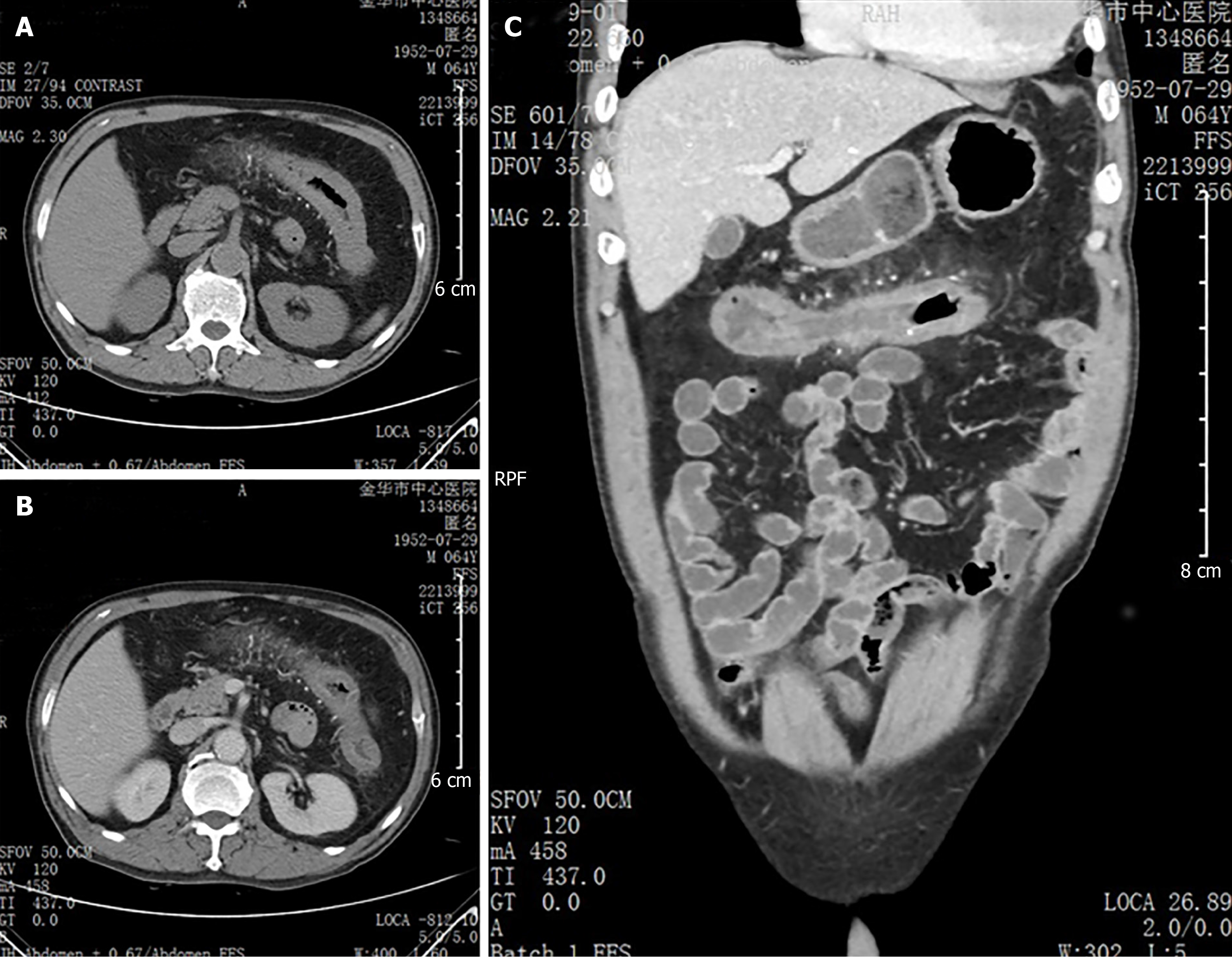
Figure 1 Contrast-enhanced abdominal computed tomography images showing colonic wall thickening and threadlike calcification of the mesenteric vein along the transverse colon.
A, B: Coronal; C: Axial.
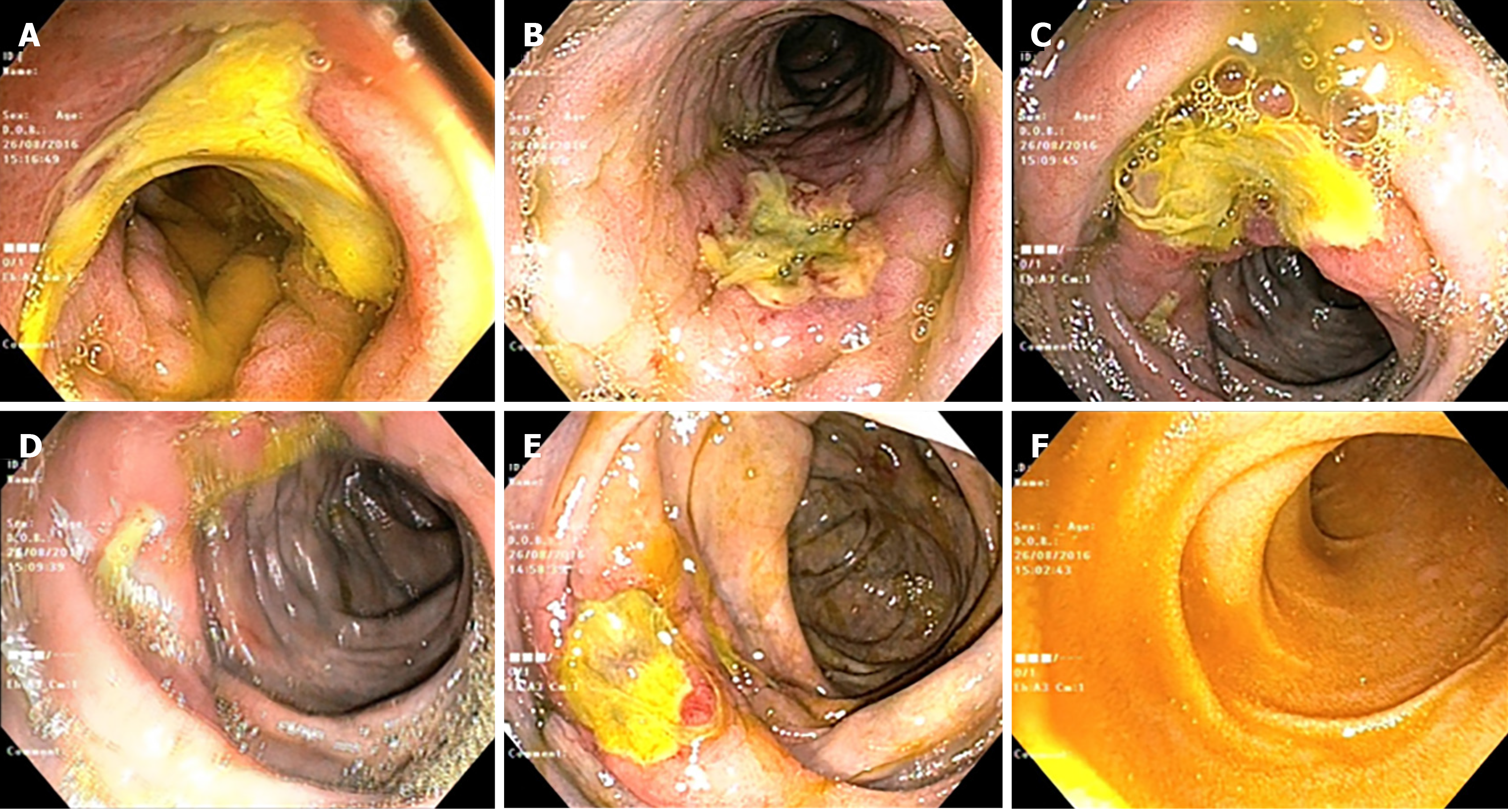
Figure 2 Colonoscopy findings.
A: Rectum; B: Descending colon; C: Transverse colon; D: Ascending colon; E: Ileocecum; F: Terminal ileum. Deep and circumferential ulcerations were observed in the descending, transverse, and ascending colon. Purple-blue mucosa was discovered in the descending colon, transverse colon, ascending colon, and ileocecum.
Figure 3 Pathological findings.
A, B: Low-power (100 ×) (A) and high-power (B) views of hematoxylin-eosin staining, showing obvious thickening and calcification of the vein walls and mucosal infiltration of eosinophils (200 ×); C: High-power view of Masson trichrome staining showing dense perivascular and mucosal collagen degeneration (200 ×).
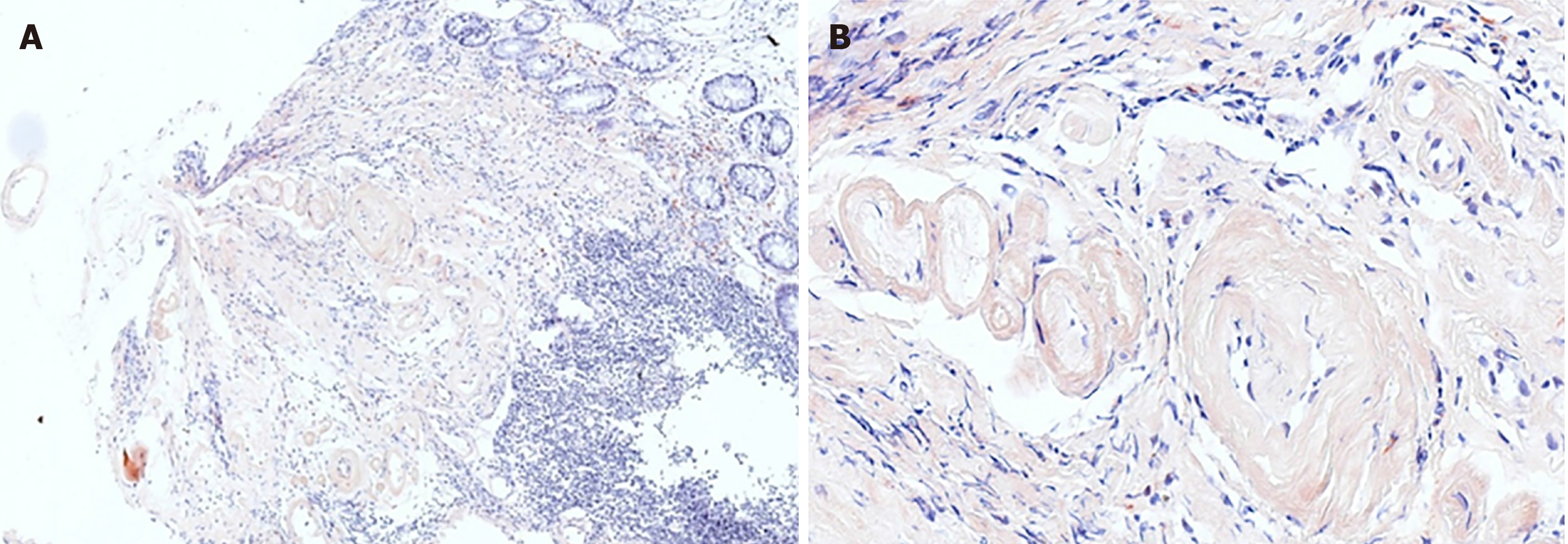
Figure 4 Pathological findings.
A, B: Low-power (100 ×) (A) and high-power views of Congo red staining showing amyloidosis of the mucosa (200 ×).
Figure 5 Disappearance of the threadlike calcification of the mesenteric vein.
A: Follow-up colonoscopy (at 3 mo) showed the remittance of ulceration in the transverse colon but the persistence of purple-blue discoloration; B: Coronal; C: Axial. Follow-up computed tomography (at 1 year) indicated the disappearance of the threadlike calcification of the mesenteric vein.
- Citation: Hu YB, Hu ML, Ding J, Wang QY, Yang XY. Mesenteric phlebosclerosis with amyloidosis in association with the long-term use of medicinal liquor: A case report. World J Clin Cases 2020; 8(4): 798-805
- URL: https://www.wjgnet.com/2307-8960/full/v8/i4/798.htm
- DOI: https://dx.doi.org/10.12998/wjcc.v8.i4.798


