Copyright
©The Author(s) 2020.
World J Clin Cases. Feb 26, 2020; 8(4): 790-797
Published online Feb 26, 2020. doi: 10.12998/wjcc.v8.i4.790
Published online Feb 26, 2020. doi: 10.12998/wjcc.v8.i4.790
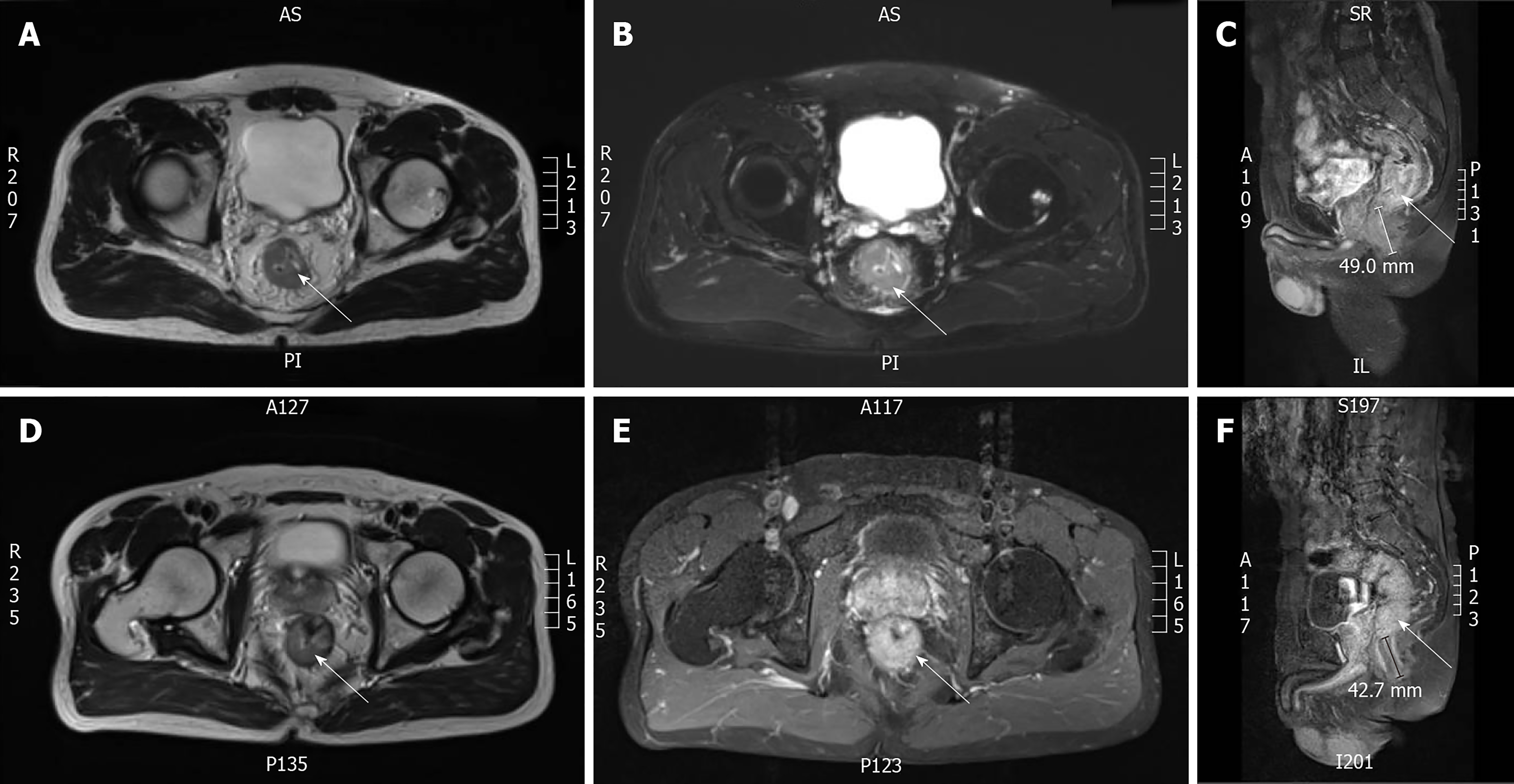
Figure 1 Preoperative pelvic magnetic resonance imaging.
A-C: Preoperative pelvic magnetic resonance imaging (MRI) of Case 1 (white arrow shows the rectal cancer, 2013/10/23); D-F: Preoperative pelvic MRI of Case 2 (white arrow shows the rectal cancer, 2015/11/05).
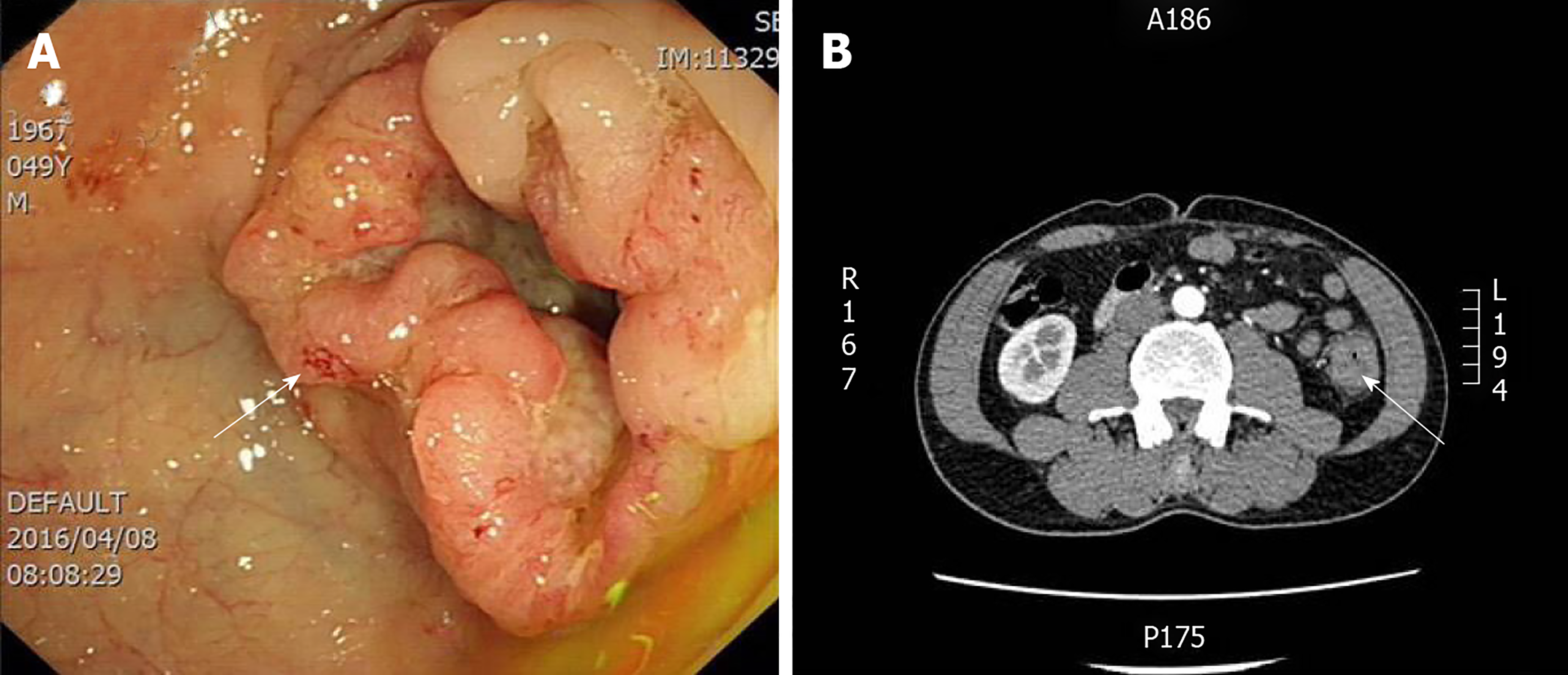
Figure 2 Preoperative performance of Case 3.
A: Preoperative colonoscopy of Case 3 (white arrow shows the colon cancer, 2016/04/08); B: Preoperative abdominal computed tomography of Case 3 (white arrow shows the colon cancer, 2016/04/11).
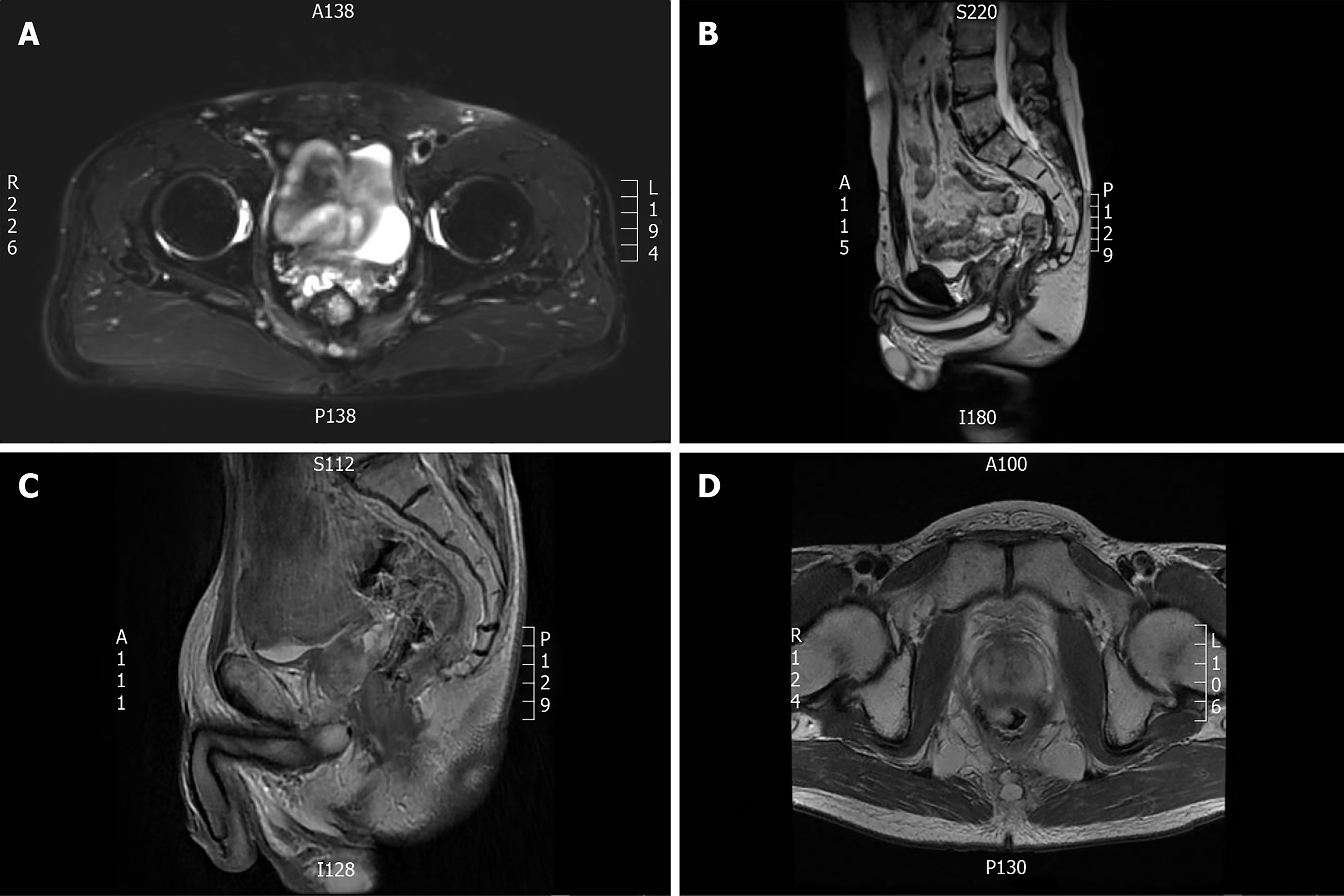
Figure 3 Recent magnetic resonance imaging reexamination.
A, B: Recent magnetic resonance imaging (MRI) reexamination of Case 1 (2018/06/04); C, D: Recent MRI reexamination of Case 2 (2017/07/19).
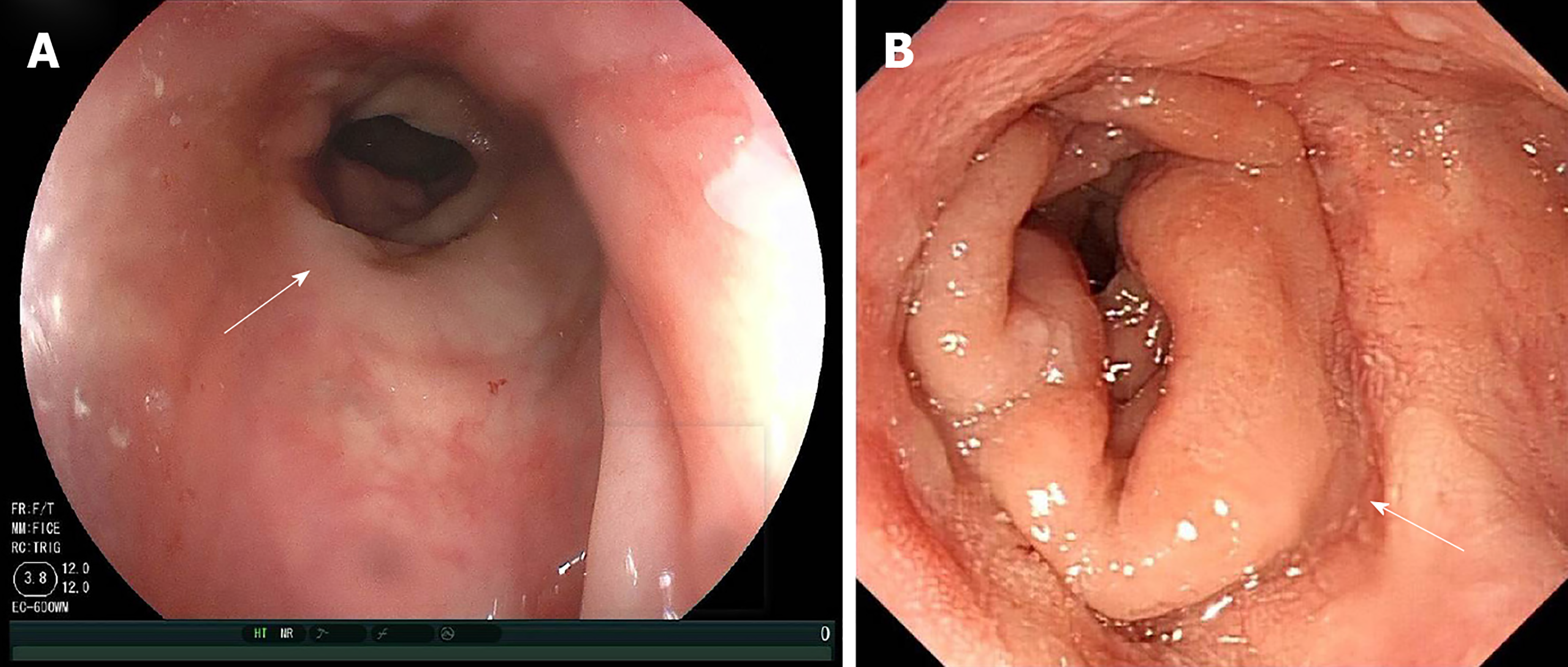
Figure 4 Recent reexamination colonoscopy.
A: Recent reexamination colonoscopy of Case 1 (white arrow shows the anastomotic stoma, 2018/12/13); B: Recent reexamination colonoscopy of Case 2 (white arrow shows the anastomotic stoma, 2018/11/19).
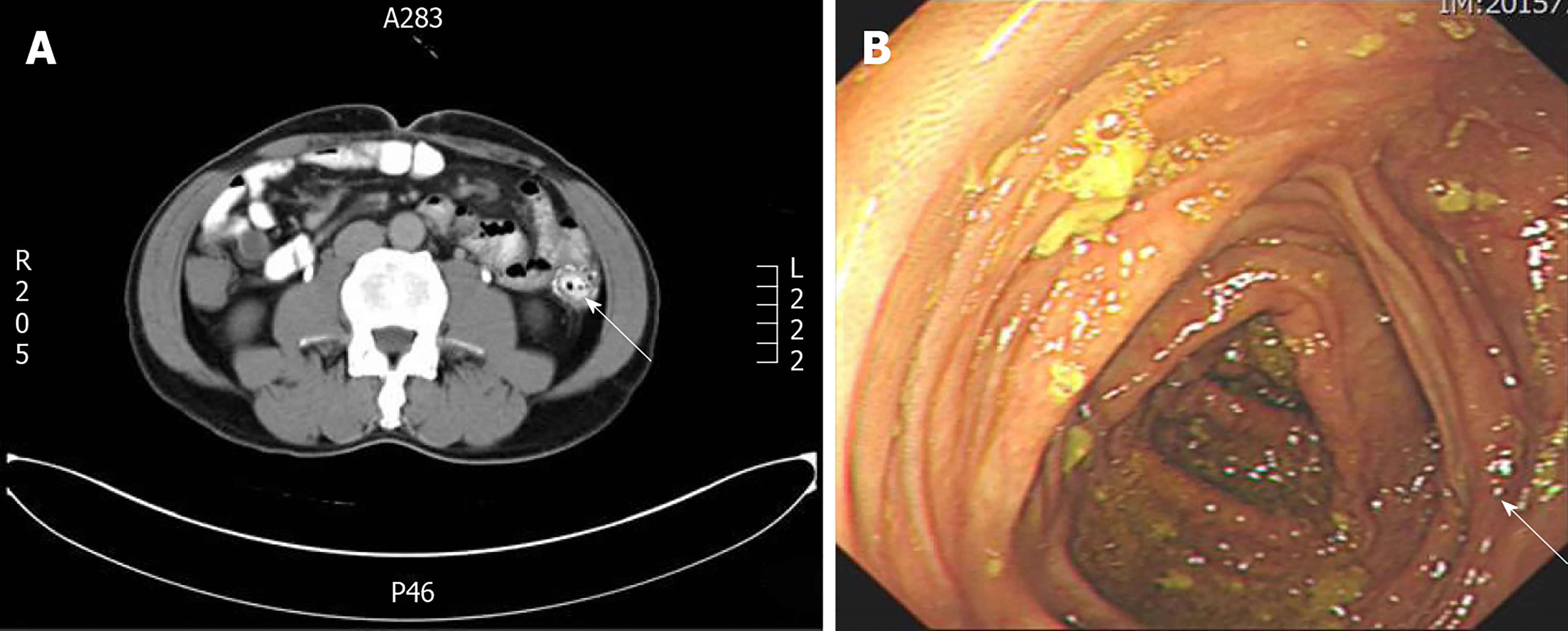
Figure 5 Recent reexamination of Case 3.
A: Recent abdominal computed tomography reexamination of Case 3 (white arrow shows the anastomotic stoma, 2018/01/03); B: Recent reexamination colonoscopy of Case 3 (white arrow shows the anastomotic stoma, 2018/01/04).
- Citation: Gu GL, Duan FX, Zhang Z, Wei XM, Cui L, Zhang B. Must pilots permanently quit flying career after treatment for colorectal cancer? - Medical waiver for Air Force pilots with colorectal cancer: Three case reports. World J Clin Cases 2020; 8(4): 790-797
- URL: https://www.wjgnet.com/2307-8960/full/v8/i4/790.htm
- DOI: https://dx.doi.org/10.12998/wjcc.v8.i4.790


