Copyright
©The Author(s) 2020.
World J Clin Cases. Jan 26, 2020; 8(2): 398-403
Published online Jan 26, 2020. doi: 10.12998/wjcc.v8.i2.398
Published online Jan 26, 2020. doi: 10.12998/wjcc.v8.i2.398
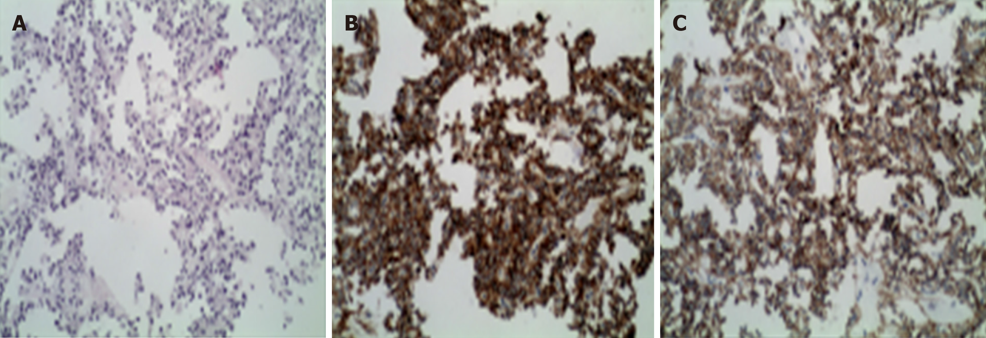
Figure 1 Pathology of solid pseudo-papillary tumor of the pancreas with liver metastasis.
A: The tumor is less heterogeneous, the cytoplasm is eosinophilic, and the tumor cells are arranged in a pseudo-nipple around the blood vessels; B: Positive cytoplasmic staining for CD10 (×200); C: Positive cytoplasmic staining for CD56 (×200).
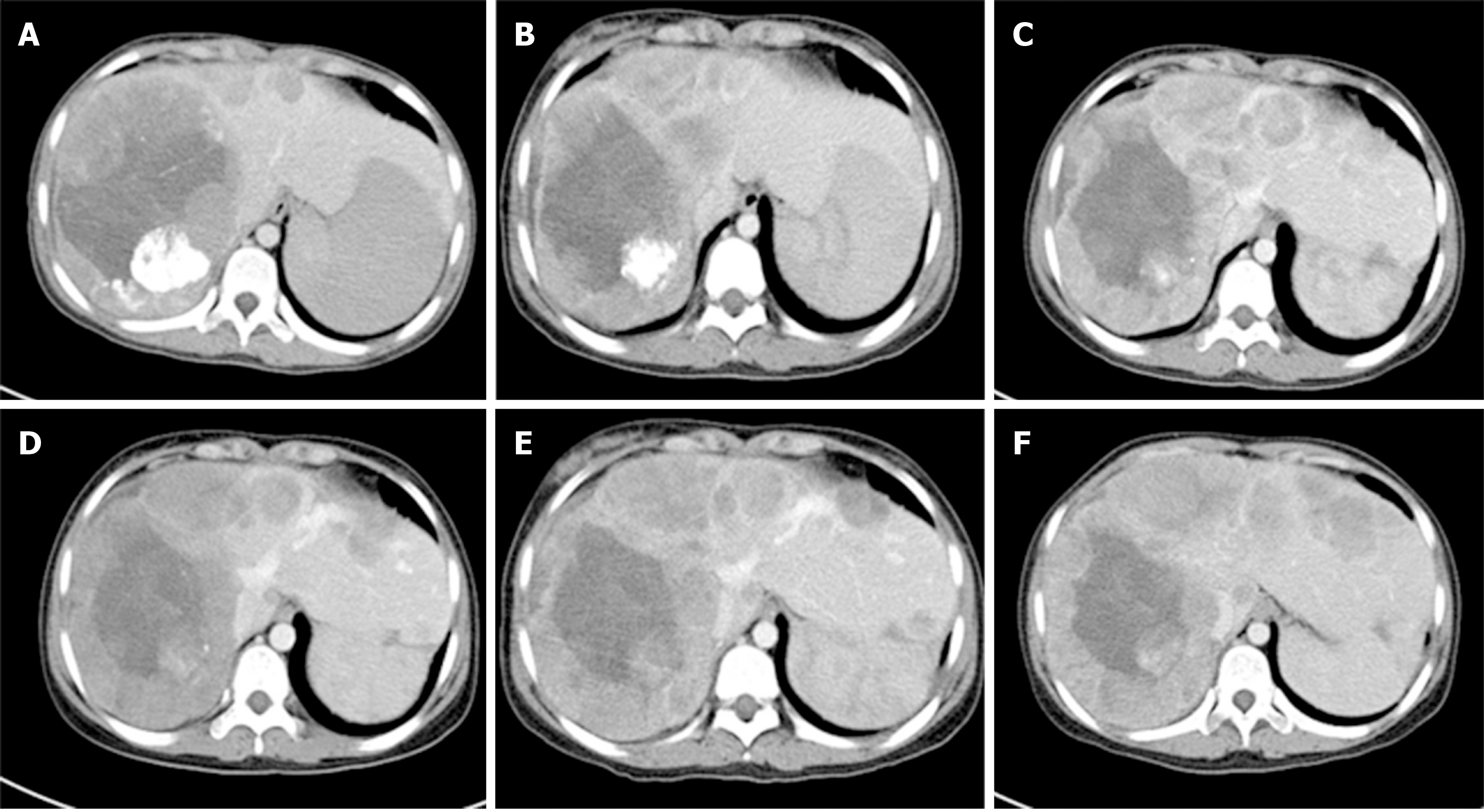
Figure 2 Computed tomography of the liver.
A: Before cryoablation, abdominal computed tomography found multiple nodules in the right lobe of the liver; B-F: Follow-up computed tomography scans at 1 year (B), 2 years (C), 3 years (D), 4 years (E), and 5 years (F).
Figure 3 T cell subsets in patient’s peripheral blood post-treatment.
A: CD3+ cell population; B: CD4+ and CD8+ cell population; C: Natural killer cell population.
- Citation: Ma YY, Chen JB, Shi JJ, Niu LZ, Xu KC. Cryoablation for liver metastasis from solid pseudopapillary tumor of the pancreas: A case report. World J Clin Cases 2020; 8(2): 398-403
- URL: https://www.wjgnet.com/2307-8960/full/v8/i2/398.htm
- DOI: https://dx.doi.org/10.12998/wjcc.v8.i2.398


