Copyright
©The Author(s) 2020.
World J Clin Cases. Jan 26, 2020; 8(2): 337-342
Published online Jan 26, 2020. doi: 10.12998/wjcc.v8.i2.337
Published online Jan 26, 2020. doi: 10.12998/wjcc.v8.i2.337
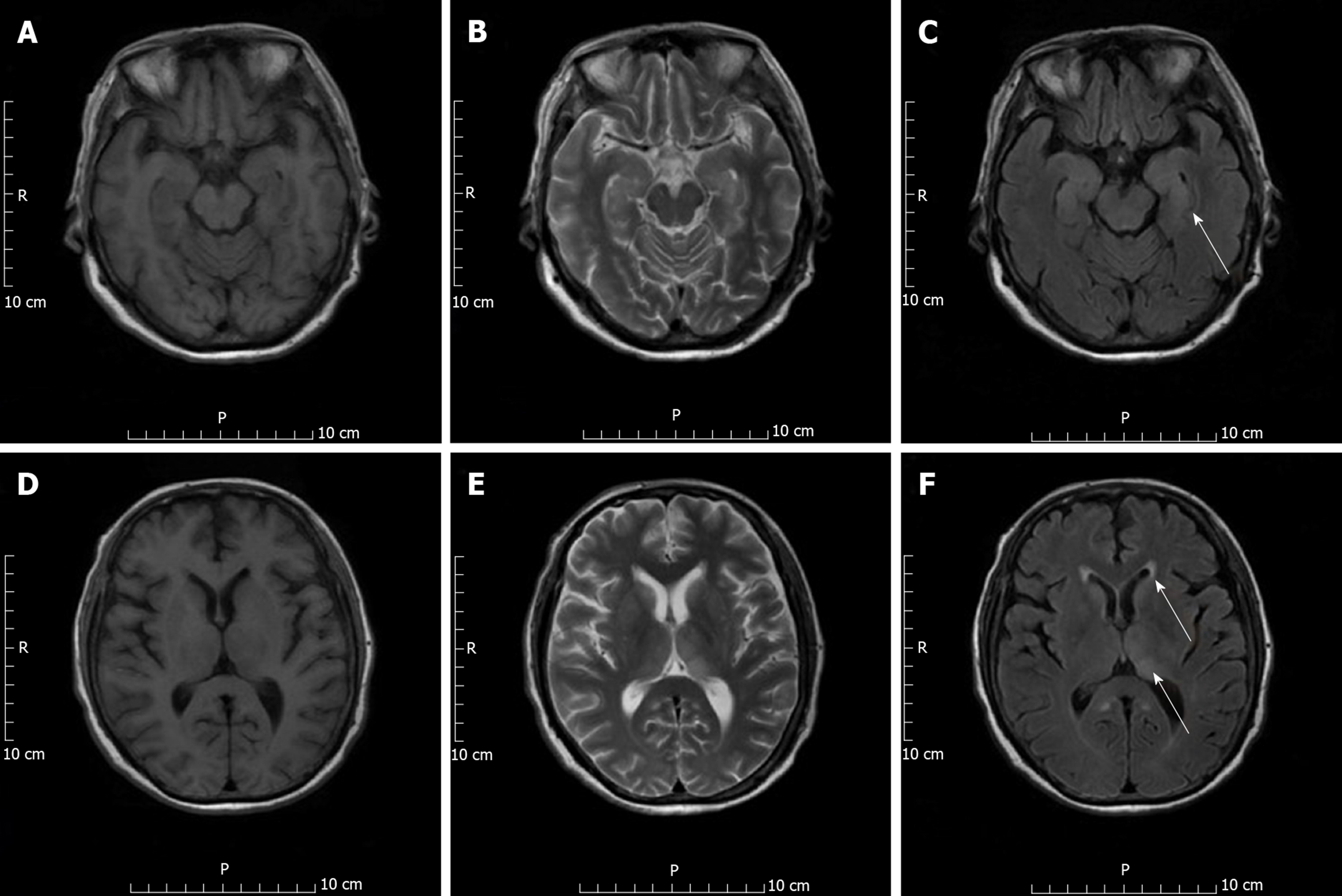
Figure 1 Enhanced magnetic resonance imaging scan of the patient’s brain on post-transplantation day 19.
A, B, D, E: Lesions can be seen in T1 image (A, D) and T2 image (B, E); C: Hyperintense lesion is seen in this T2 fluid attenuated inversion recovery image in parahippocampal gyrus; F: Hyperintense lesions are seen in this T2 fluid attenuated inversion recovery image in the bilateral thalamus and caudate nucleus. Arrows indicate the lesions.
- Citation: Qi ZL, Sun LY, Bai J, Zhuang HZ, Duan ML. Japanese encephalitis following liver transplantation: A rare case report. World J Clin Cases 2020; 8(2): 337-342
- URL: https://www.wjgnet.com/2307-8960/full/v8/i2/337.htm
- DOI: https://dx.doi.org/10.12998/wjcc.v8.i2.337


