Copyright
©The Author(s) 2020.
World J Clin Cases. Sep 26, 2020; 8(18): 4228-4233
Published online Sep 26, 2020. doi: 10.12998/wjcc.v8.i18.4228
Published online Sep 26, 2020. doi: 10.12998/wjcc.v8.i18.4228
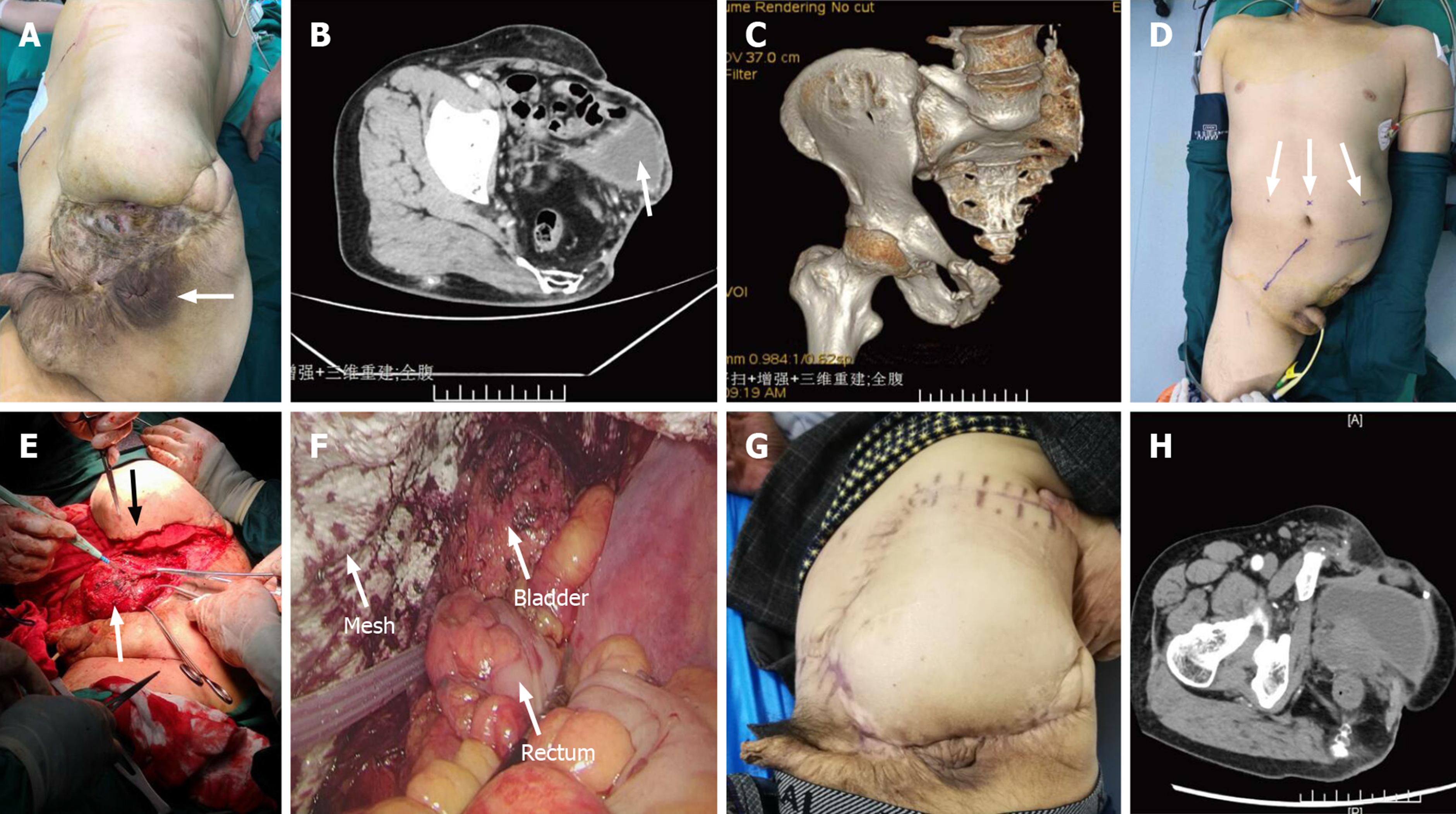
Figure 1 Physical and imaging examinations.
A: Physical examination showed the position of the anus (arrow), a perineal bulge and festering skin; B: Contrast-enhanced computed tomography revealed that a part of the small intestine and the bladder (arrow) were slightly protruding from the bottom of the pelvis; C: Computed tomography three-dimensional imaging showed the lack of pelvic structures; D: We marked the location of trocars and supplying blood vessels of the flap with ultrasound before operation; E: The perineal approach allowed us to dissociate the bladder (white arrow), and skin flap transfer (black arrow) was performed; F: The laparoscopic field of vision clearly demonstrated the positional relationship between the mesh, bladder, and rectum in stage 3; G and H: No signs of recurrence were found through general physical examination or computed tomography 2 yr after the surgery.
- Citation: Chen K, Lan YZ, Li J, Xiang YY, Zeng DZ. Mine disaster survivor’s pelvic floor hernia treated with laparoscopic surgery and a perineal approach: A case report. World J Clin Cases 2020; 8(18): 4228-4233
- URL: https://www.wjgnet.com/2307-8960/full/v8/i18/4228.htm
- DOI: https://dx.doi.org/10.12998/wjcc.v8.i18.4228


