Copyright
©The Author(s) 2020.
World J Clin Cases. Sep 26, 2020; 8(18): 4122-4127
Published online Sep 26, 2020. doi: 10.12998/wjcc.v8.i18.4122
Published online Sep 26, 2020. doi: 10.12998/wjcc.v8.i18.4122
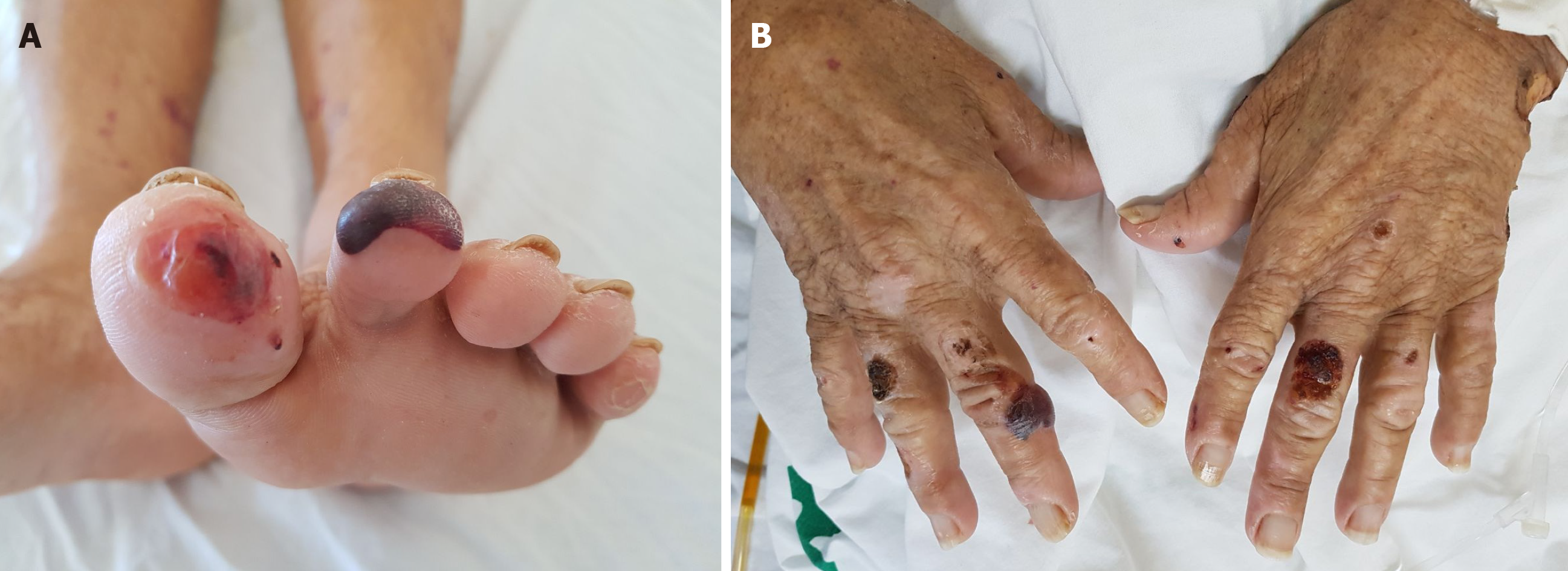
Figure 1 Blistering skin lesions suggesting pemphigus on foot (A) and hands (B).
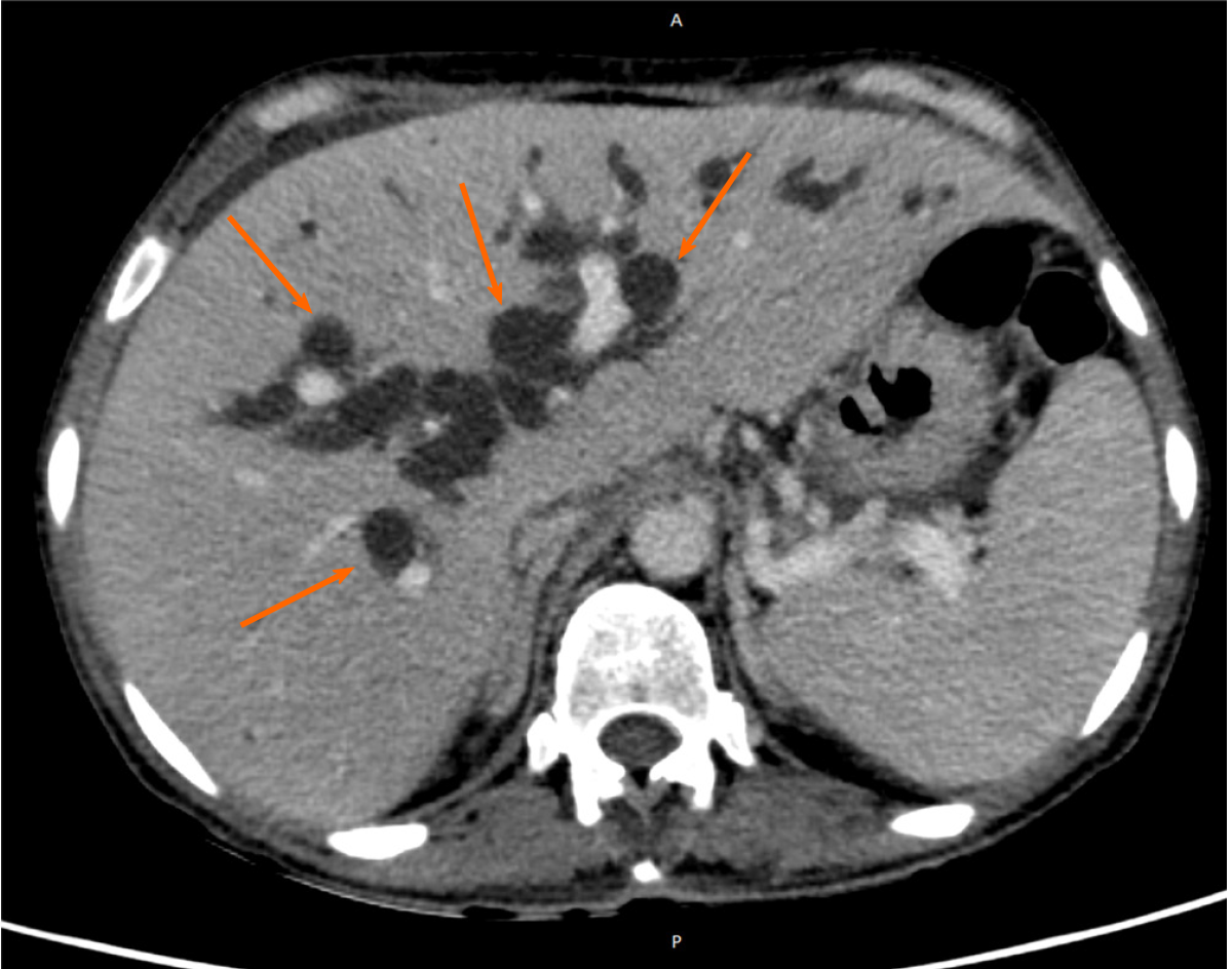
Figure 2 Axial plane image of computed tomography obtained in the portal phase showing significant intrahepatic biliary tract dilatation (orange arrows).
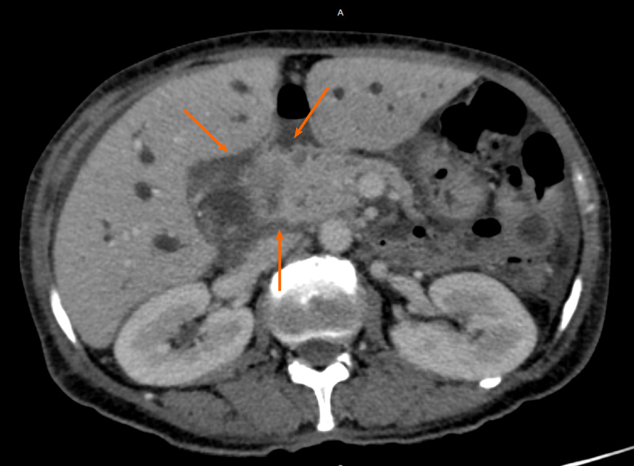
Figure 3 Axial plane image of computed tomography obtained in the portal phase showing an infiltrative aspect of the tumor, with irregular contour and mild contrast enhancement located at the common bile duct (orange arrows) determining biliary tract dilatation.
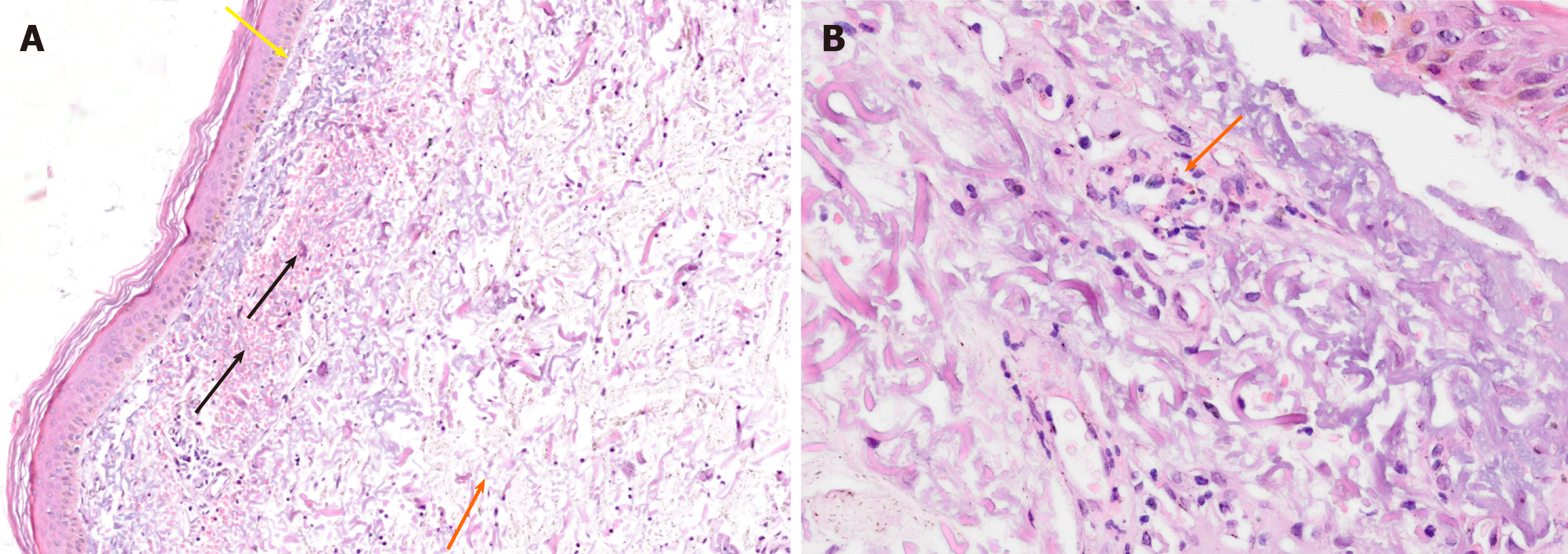
Figure 4 Skin lesions biopsy stained with hematoxylin-eosin showed elastosis (yellow arrow), edema (orange arrow), bleeding focus and red cells extravasation (black arrows) (A) and capillary destruction (B).
Figure 5 Timeline of events reported in the clinical case.
- Citation: Lemaire CC, Portilho ALC, Pinheiro LV, Vivas RA, Britto M, Montenegro M, Rodrigues LFF, Arruda S, Lyra AC, Cavalcante LN. Sweet syndrome as a paraneoplastic manifestation of cholangiocarcinoma: A case report. World J Clin Cases 2020; 8(18): 4122-4127
- URL: https://www.wjgnet.com/2307-8960/full/v8/i18/4122.htm
- DOI: https://dx.doi.org/10.12998/wjcc.v8.i18.4122


