Copyright
©The Author(s) 2020.
World J Clin Cases. Jul 26, 2020; 8(14): 3082-3089
Published online Jul 26, 2020. doi: 10.12998/wjcc.v8.i14.3082
Published online Jul 26, 2020. doi: 10.12998/wjcc.v8.i14.3082
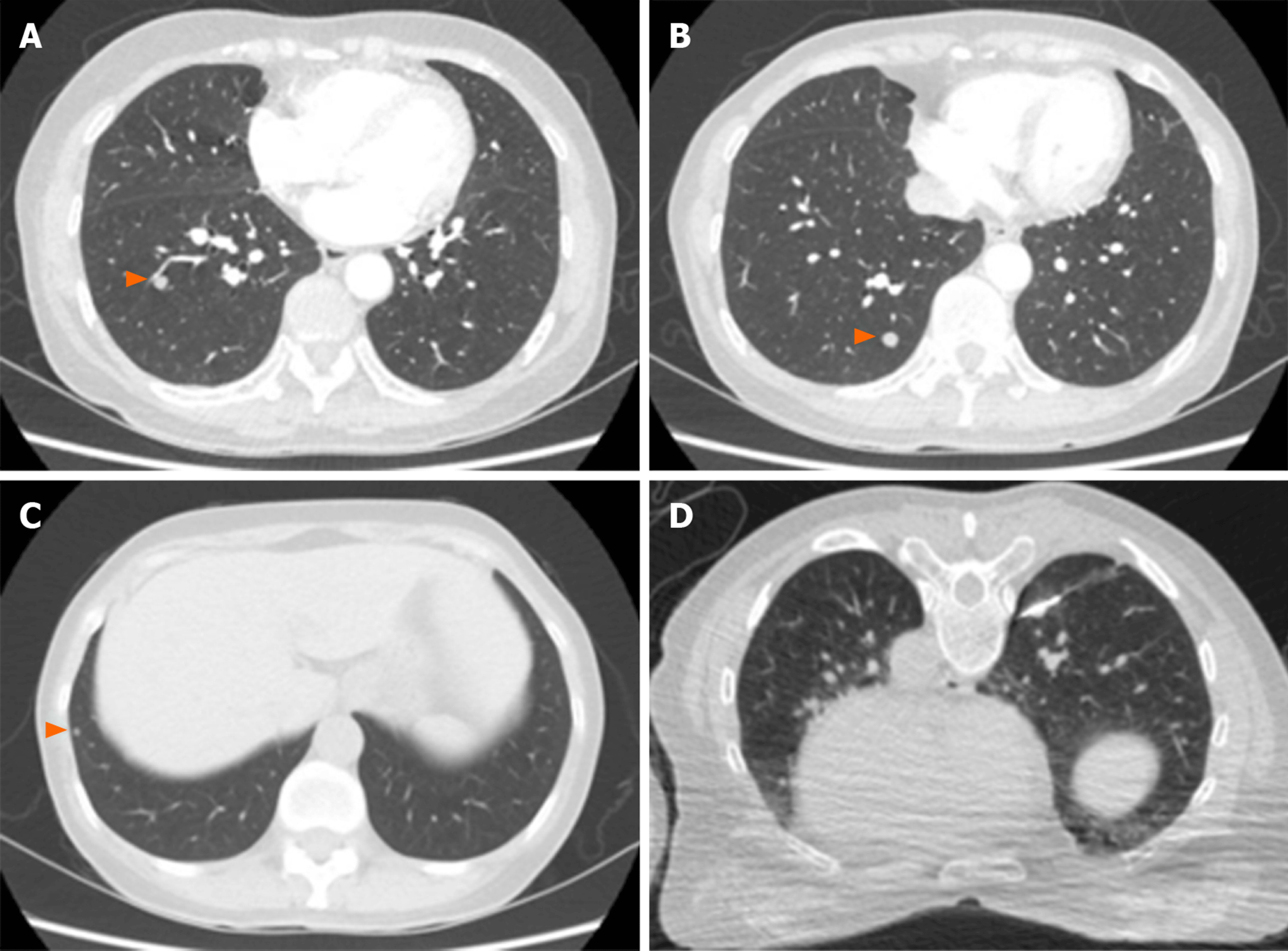
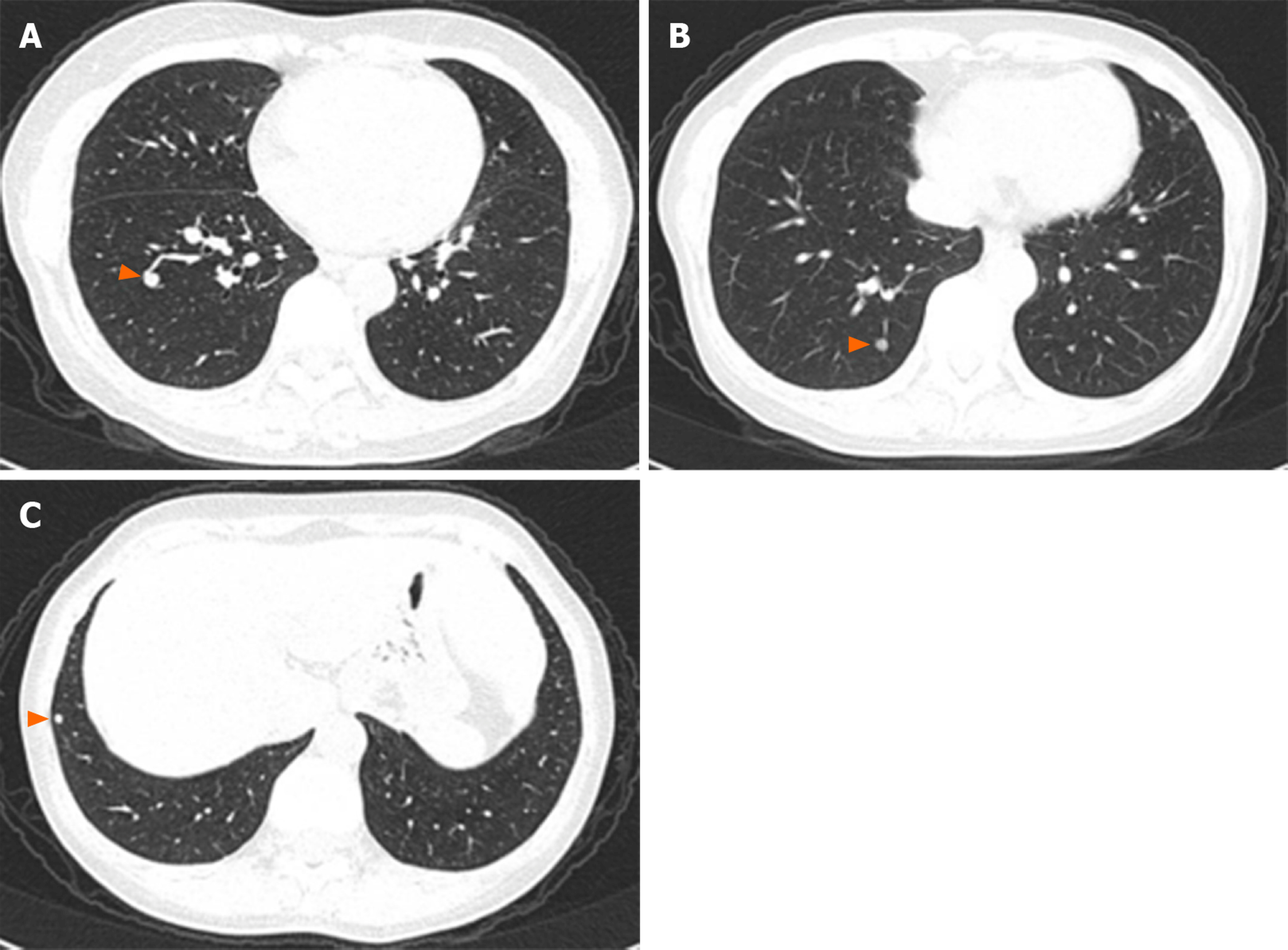
Figure 1 Chest high-resolution computed tomography imaging.
A-C: The three largest lung nodules on chest high-resolution computed tomography; D: Computed tomography-guided percutaneous lung puncture biopsy of the largest nodule in the right lower lobe.
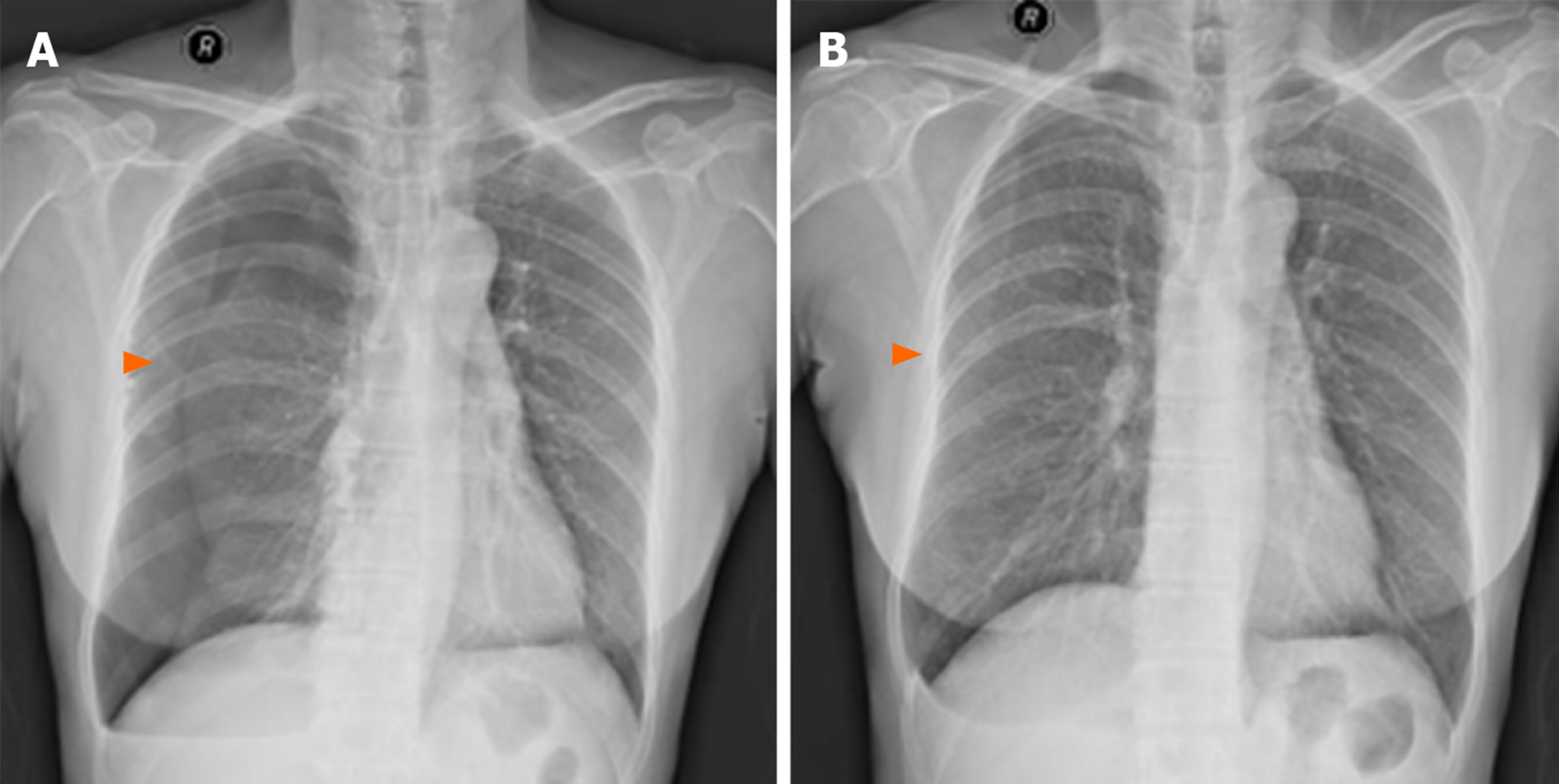
Figure 2 Chest X-ray imaging.
A: Complication of pneumothorax after computed tomography-guided percutaneous lung puncture biopsy; B: The patient recovered after thoracic puncture and aspiration.
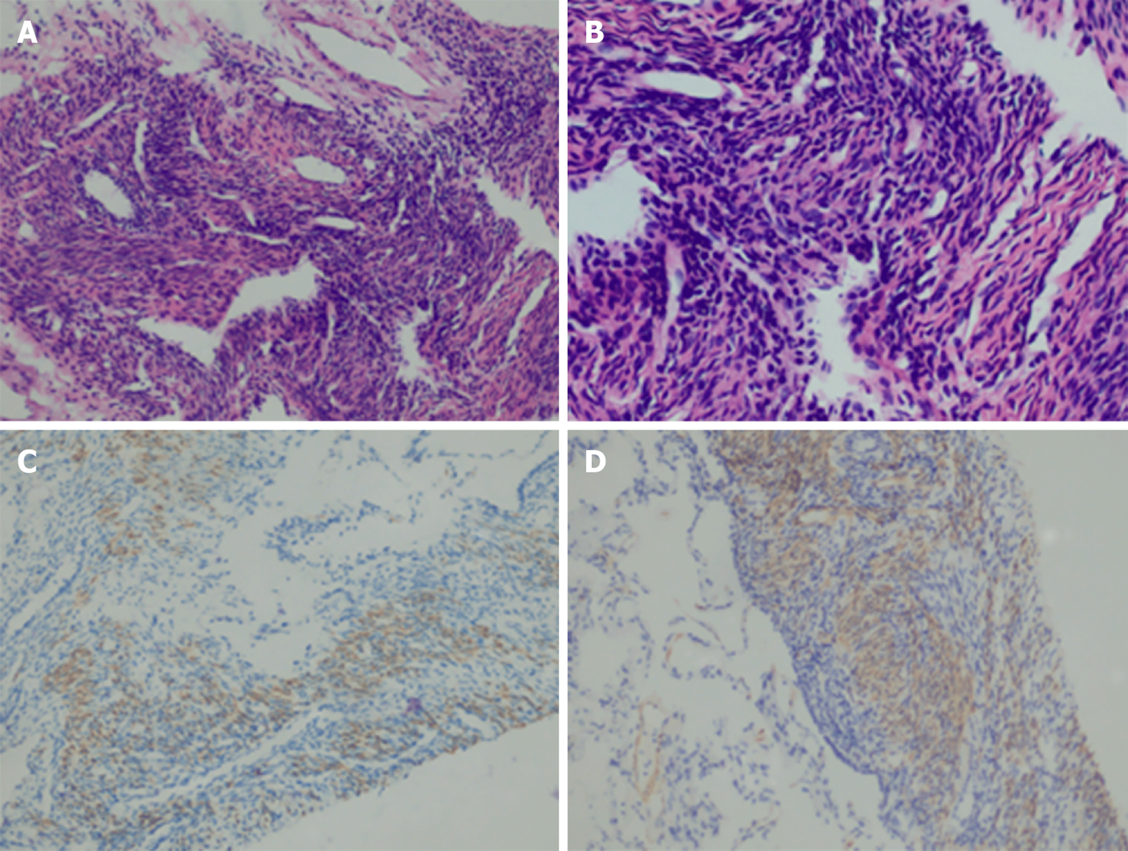
Figure 3 Histological findings.
A-D: Histopathological examination of the lung puncture biopsy specimen: A few spindle cells were seen, and a spindle cell tumor was considered; immunohistochemical smooth-muscle labeling revealed Desmin(+) and SMA(+), suggesting smooth muscle origin.
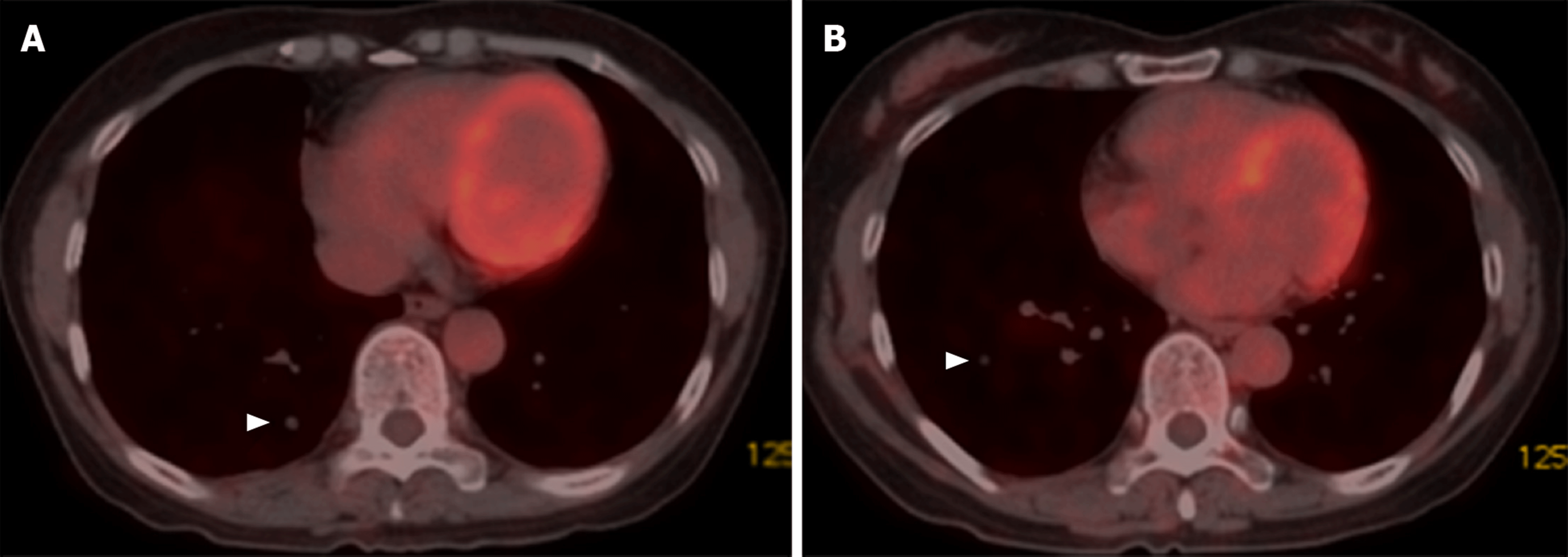
Figure 4 Positron emission tomography-computed tomography imaging.
A and B: Positron emission tomography-computed tomography showed multiple nodules in both lungs with no uptake of fluorodeoxyglucose.
Figure 5 Follow-up with chest high-resolution computed tomography imaging after 6 mo.
A-C: No progression.
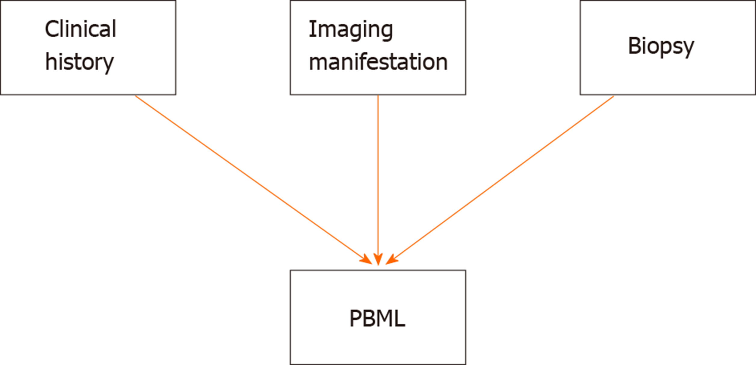
Figure 6 Three major diagnostic aspects for pulmonary benign metastatic leiomyoma.
PBML: Pulmonary benign metastatic leiomyoma.
- Citation: Dai HY, Guo SL, Shen J, Yang L. Pulmonary benign metastasizing leiomyoma: A case report and review of the literature. World J Clin Cases 2020; 8(14): 3082-3089
- URL: https://www.wjgnet.com/2307-8960/full/v8/i14/3082.htm
- DOI: https://dx.doi.org/10.12998/wjcc.v8.i14.3082


