Copyright
©The Author(s) 2020.
World J Clin Cases. Jul 26, 2020; 8(14): 3031-3038
Published online Jul 26, 2020. doi: 10.12998/wjcc.v8.i14.3031
Published online Jul 26, 2020. doi: 10.12998/wjcc.v8.i14.3031
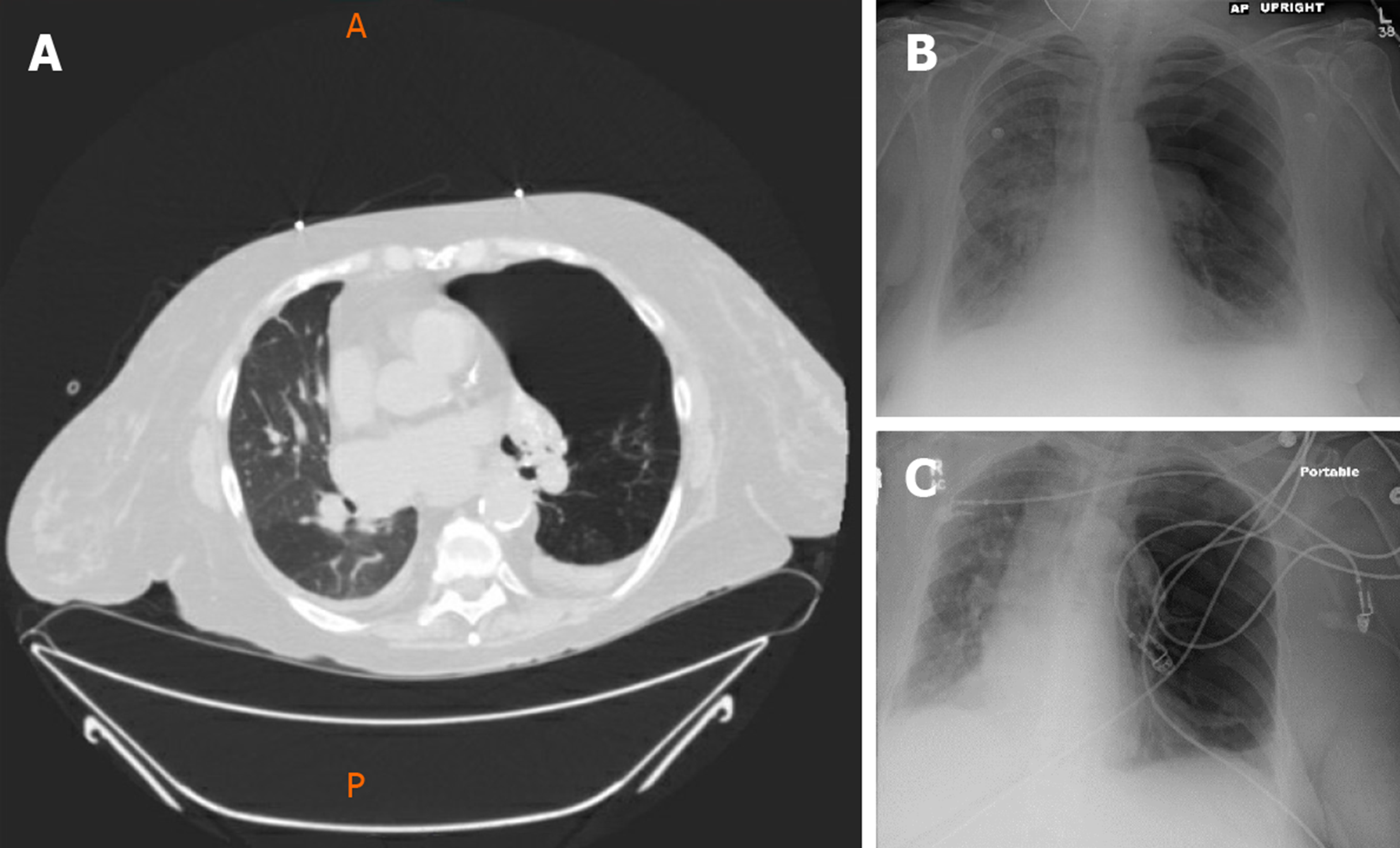
Figure 1 Computed tomography (prone position) of the chest (A) and chest radiographs AP upright (B) and portable (C), show native left lung and transplanted right lung.
There is a large bulla in the left upper lobe. Note subtle mosaicism of the transplanted right lung allograft on computed tomography.
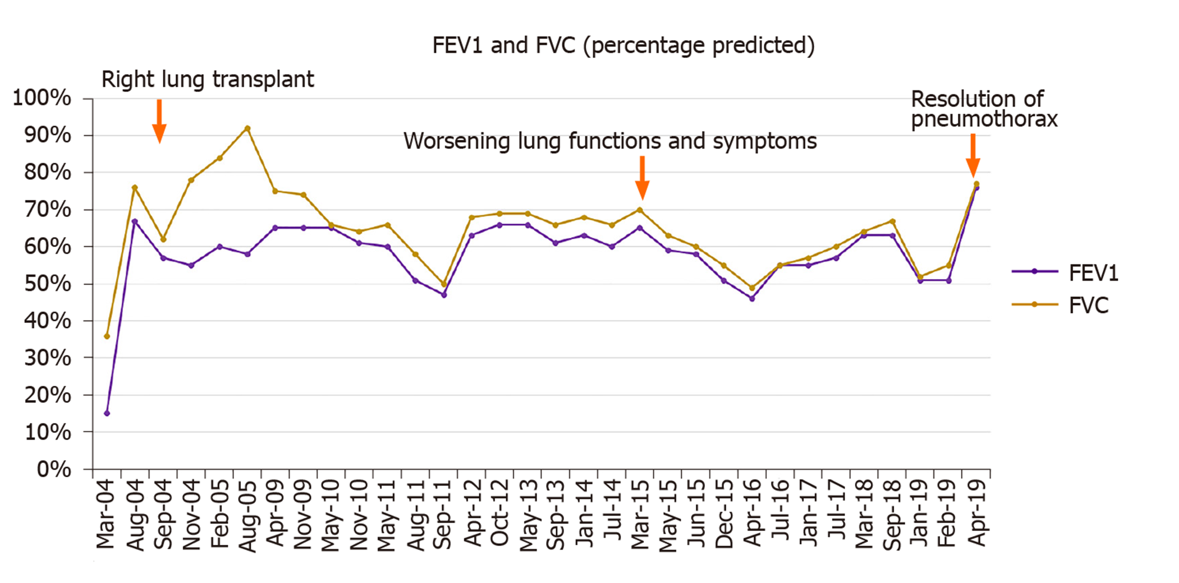
Figure 2 Linear graph demonstrating the FEV1 and FVC (percentage predicted) for the patient on follow-ups.
Note improvement in lung functions after right lung transplantation (Aug 2004) and subsequent decline in 2015 followed by improvement in 2019 on the resolution of pneumothorax.
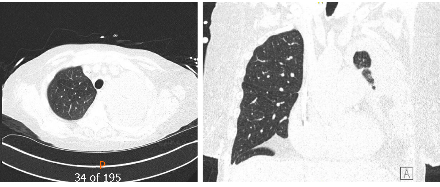
Figure 3 Computed tomography chest (Axial and coronal reformatted images) show a large left pleural effusion with near-complete opacification of the left lung parenchyma.
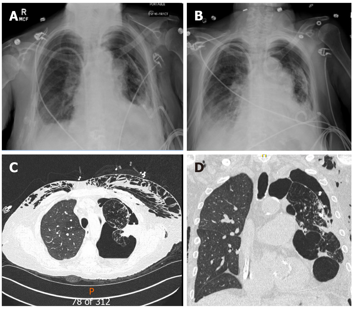
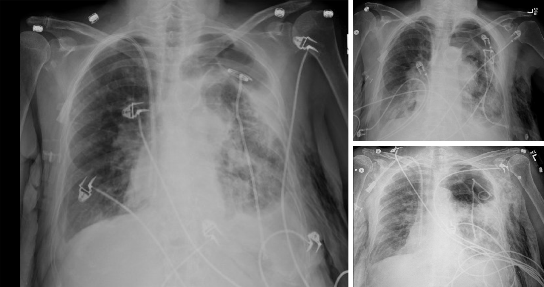
Figure 4 The native left lung is partially collapsed, there is subcutaneous emphysema in the left chest wall.
A and B: Portable chest radiographs; C and D: Computed tomography chest (axial and coronal reformation) images reveal a large left hydropneumothorax with an indwelling pigtail drainage catheter in the left pleural space.
Figure 5 Serial portable chest radiographs show replacement of a left pleural drain within a large left hydropneumothorax with compressive atelectasis of portions of the native left lung.
Subcutaneous emphysema is again noted within the left chest wall. The transplanted right lung remains well inflated.
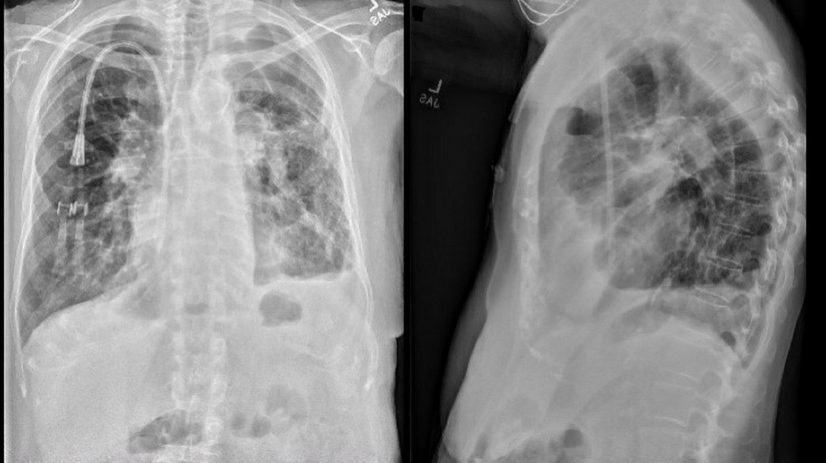
Figure 6 Chest radiographs (PA and lateral views) demonstrate auto-deflation of large left upper lobe bulla resulting in subsequent re-expansion of the previously atelectatic left lung as well as improved aeration of the transplanted right lung.
- Citation: Deshwal H, Ghosh S, Hogan K, Akindipe O, Lane CR, Mehta AC. Spontaneous pneumothorax in a single lung transplant recipient-a blessing in disguise: A case report. World J Clin Cases 2020; 8(14): 3031-3038
- URL: https://www.wjgnet.com/2307-8960/full/v8/i14/3031.htm
- DOI: https://dx.doi.org/10.12998/wjcc.v8.i14.3031


