Copyright
©The Author(s) 2020.
World J Clin Cases. Jul 6, 2020; 8(13): 2679-2702
Published online Jul 6, 2020. doi: 10.12998/wjcc.v8.i13.2679
Published online Jul 6, 2020. doi: 10.12998/wjcc.v8.i13.2679
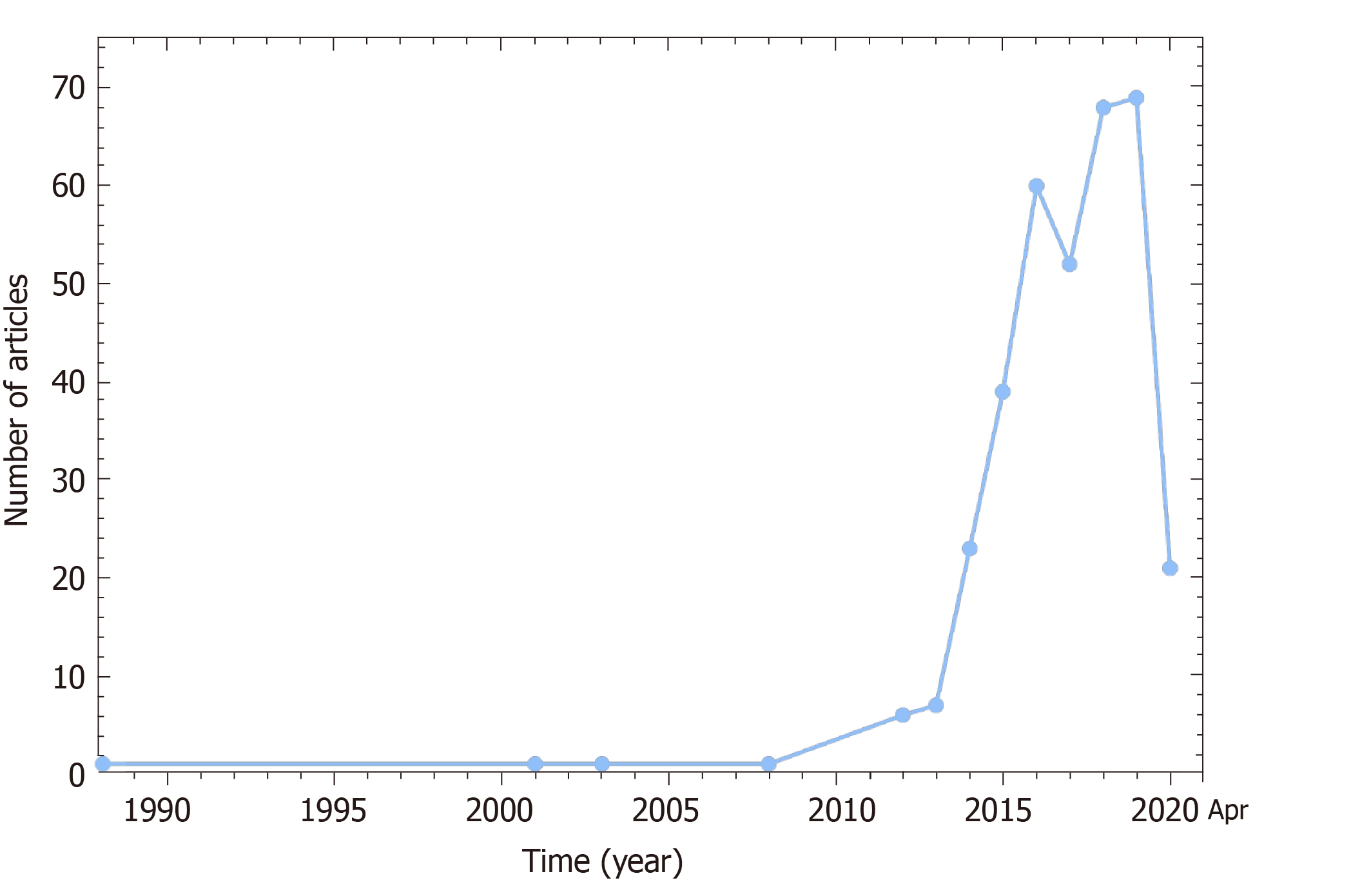
Figure 1 Article publishing trend displaying a booming development of balloon pulmonary angioplasty in chronic thromboembolic pulmonary hypertension from 1988 to April 26, 2020 (PubMed indexed).
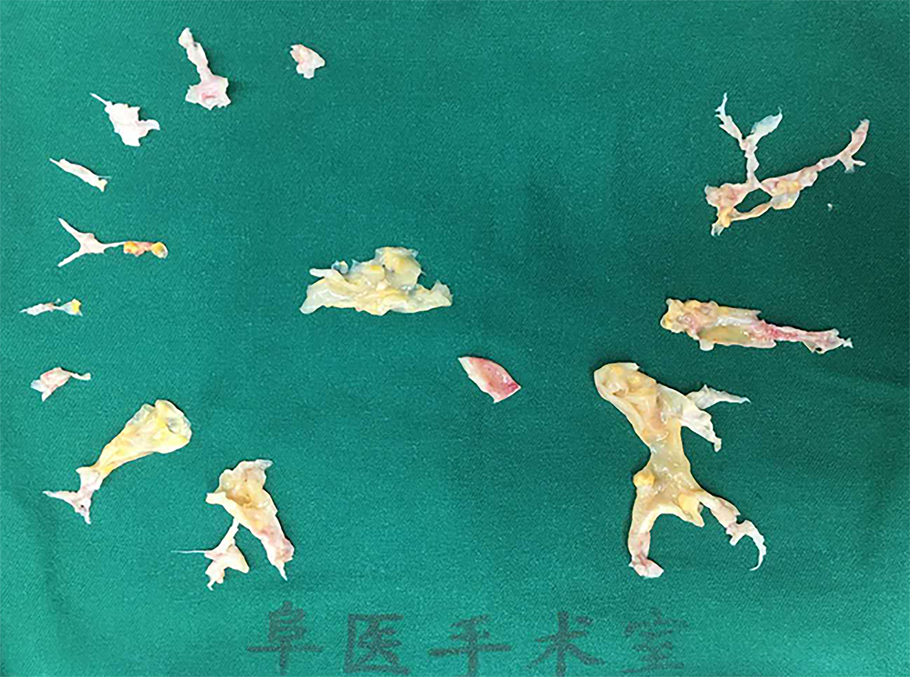
Figure 2 Endarterectomized tissues from a chronic thromboembolic pulmonary hypertension patient whose mean pulmonary arterial pressure decreased from 43 to 23 mmHg after endarterectomy.
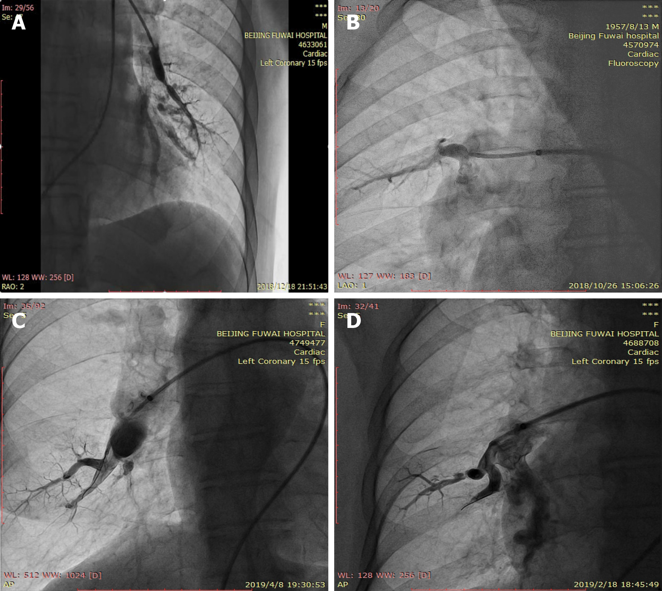
Figure 3 Angiographic classification of vessel lesions.
A: Ring-like stenosis lesion (band lesion); B: Web lesion; C: Subtotal occlusion lesion; D: Total occlusion lesion (pouch defect).
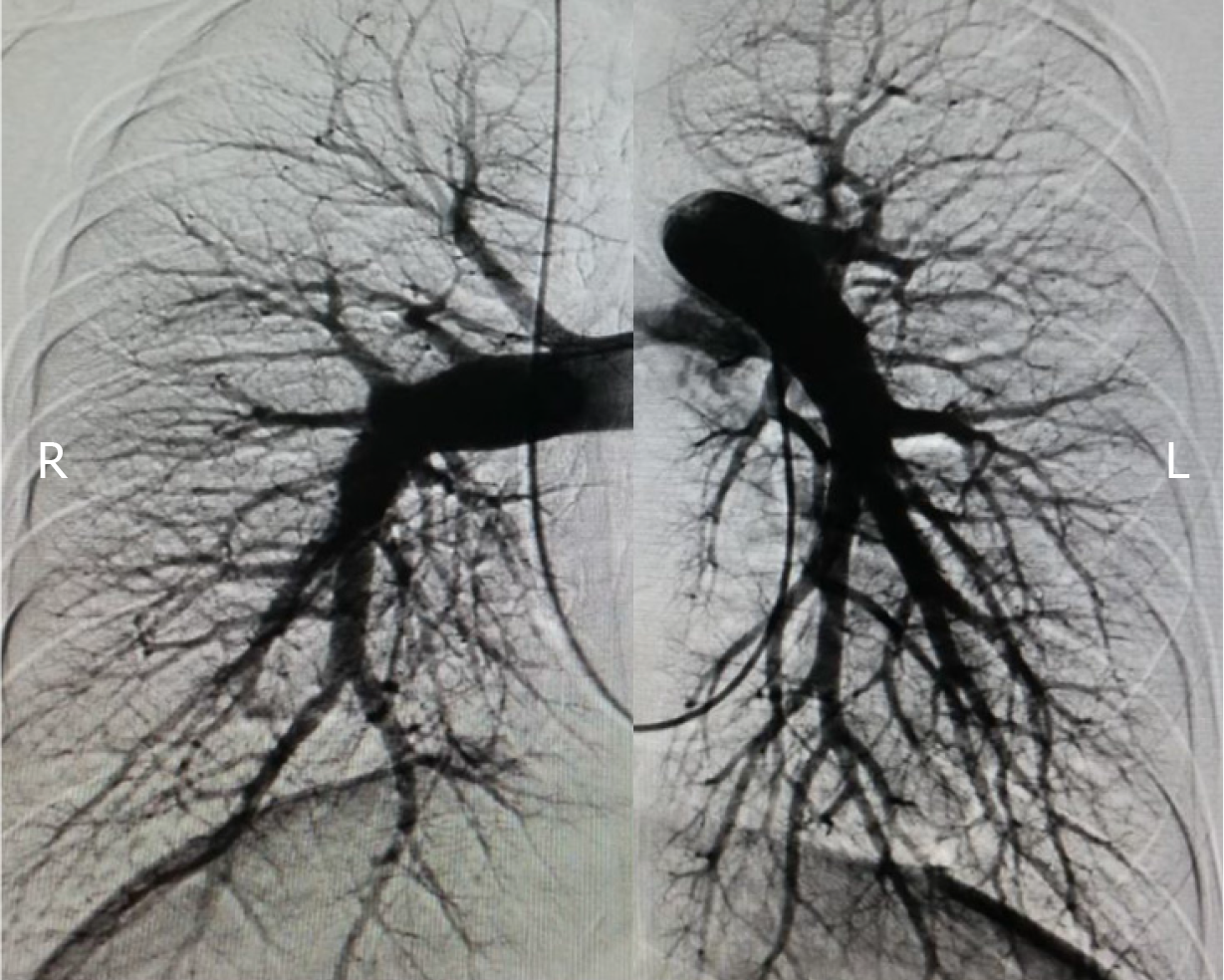
Figure 4 Non-selective pulmonary angiography of bilateral pulmonary arteries.
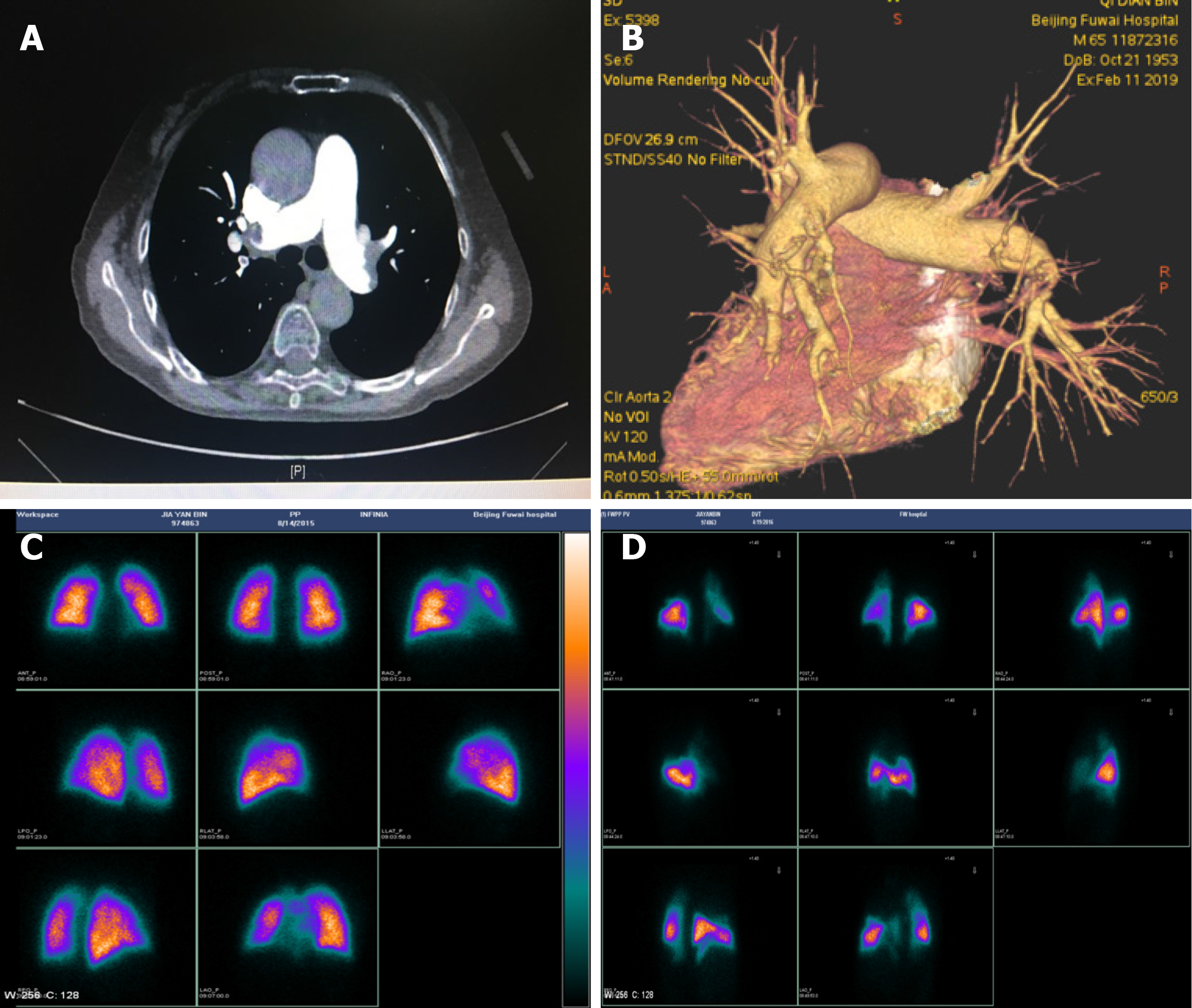
Figure 5 Computed tomographic pulmonary angiography and pulmonary perfusion scintigraphy.
A and B: Thrombi in the left and right pulmonary arteries by computed tomographic pulmonary angiography; C: Normal lung perfusion; D: Abnormal lung perfusion indicating multiple perfusion defects.
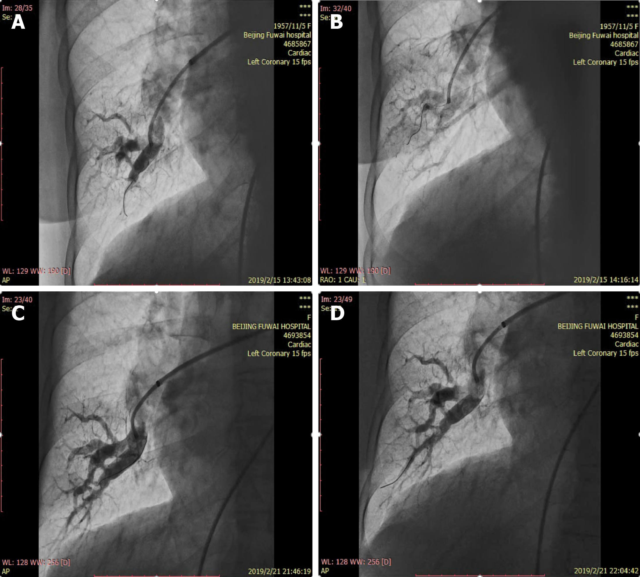
Figure 6 Representative angiographic images in the right lower lobe.
A: Subtotal occlusion lesion before the first balloon pulmonary angioplasty (BPA); B: Improved blood flow with visible venous return after the first BPA; C: Spontaneously dilated vessels with increased blood flow before the second BPA; D: Increased blood flow after the second BPA.
- Citation: Jin Q, Zhao ZH, Luo Q, Zhao Q, Yan L, Zhang Y, Li X, Yang T, Zeng QX, Xiong CM, Liu ZH. Balloon pulmonary angioplasty for chronic thromboembolic pulmonary hypertension: State of the art. World J Clin Cases 2020; 8(13): 2679-2702
- URL: https://www.wjgnet.com/2307-8960/full/v8/i13/2679.htm
- DOI: https://dx.doi.org/10.12998/wjcc.v8.i13.2679


