Copyright
©The Author(s) 2020.
World J Clin Cases. Jan 6, 2020; 8(1): 149-156
Published online Jan 6, 2020. doi: 10.12998/wjcc.v8.i1.149
Published online Jan 6, 2020. doi: 10.12998/wjcc.v8.i1.149
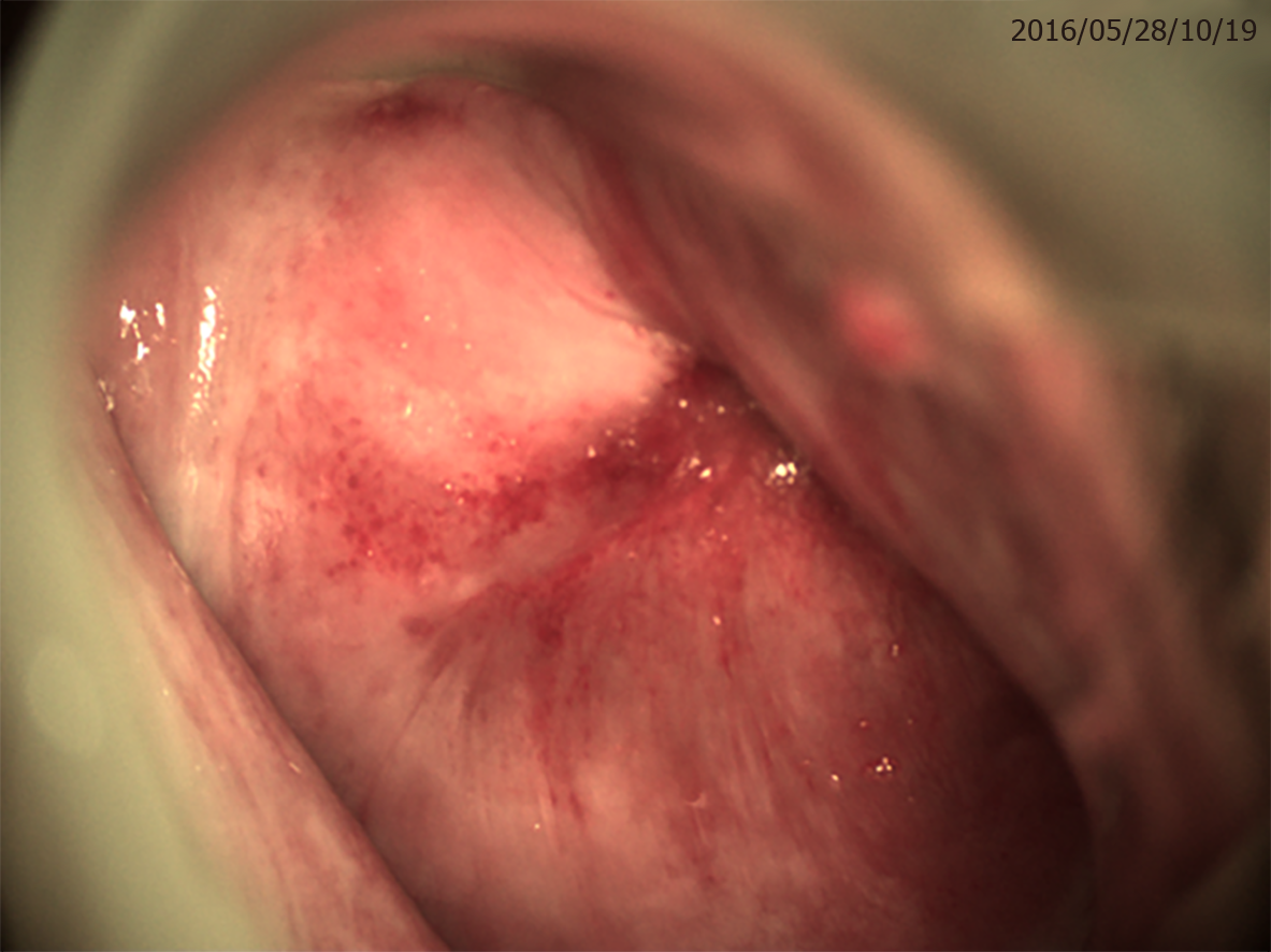
Figure 1 No distinct acetowhite lesion or anomalous vessel was detected by colposcopy, and there was no exact cervical inner orifice.
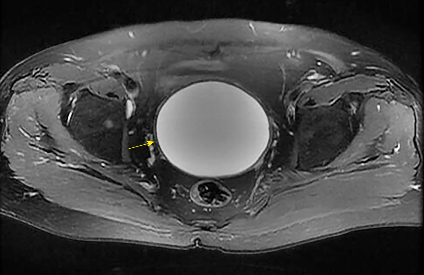
Figure 2 A large mass (yellow arrow) located in the pelvis was detected by magnetic resonance imaging.
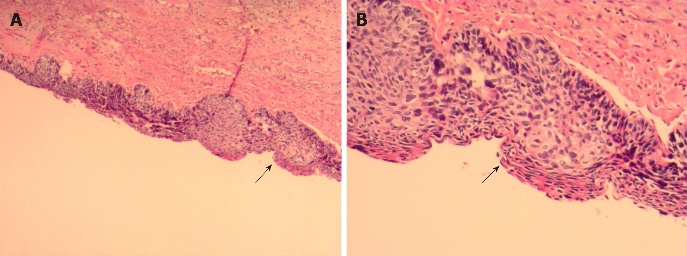
Figure 3 Pathological images.
A and B: Final pathology confirmed a large inflammatory cyst coated with a cervical high-grade squamous intraepithelial lesion (black arrows; haematoxylin and eosin staining; original magnification, ×40 for A and ×100 for B).
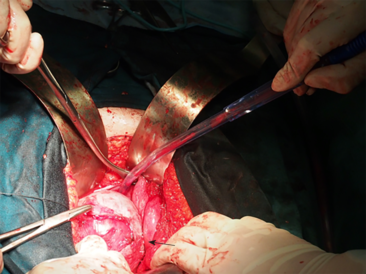
Figure 4 Operative view.
The mass was adherent with peripheric tissue (black arrow).
- Citation: Zhang K, Jiang JH, Hu JL, Liu YL, Zhang XH, Wang YM, Xue FX. Large pelvic mass arising from the cervical stump: A case report. World J Clin Cases 2020; 8(1): 149-156
- URL: https://www.wjgnet.com/2307-8960/full/v8/i1/149.htm
- DOI: https://dx.doi.org/10.12998/wjcc.v8.i1.149


